

Labelling Review row-Online
Labelling Review row-Online
Labelling Review row-Online

You also want an ePaper? Increase the reach of your titles
YUMPU automatically turns print PDFs into web optimized ePapers that Google loves.
Primary Antibodies<br />
Dysferlin<br />
Clone Ham1/7B6<br />
1 mL, 0.1 mL lyophilized NCL-Hamlet FPW<br />
Clone Ham3/17B2<br />
1 mL, 0.1 mL lyophilized NCL-Hamlet-2 F P (HIER) W<br />
Dysferlin is the protein product of the 2p13 gene that is defective in<br />
individuals with Limb-Girdle Muscular Dystrophy type 2B (LGMD2B) and<br />
Miyoshi Myopathy (MM). Dysferlin is normally localized to the muscle<br />
plasma membrane. In individuals with LGMD2B and MM, immunoreactivity<br />
to dysferlin is reported to be severely reduced or lost, depending on the type<br />
of mutation. Individuals with other neuromuscular conditions demonstrate<br />
normal labeling patterns.<br />
Product Specific Information<br />
NCL-Hamlet may require heat induced epitope retrieval in some cases.<br />
labeling with an antibody to beta-spectrin, to monitor membrane integrity, is<br />
an essential immunohistochemical control.<br />
Dystrophin Antibodies<br />
Clone Dy4/6D3<br />
2.5 mL, 1 mL lyophilized Dystrophin (Rod Domain)<br />
NCL-DYS1 FWE<br />
Clone Dy8/6C5<br />
2.5 mL, 1 mL lyophilized Dystrophin (C-terminus)<br />
NCL-DYS2 FWE<br />
Clone Dy10/12B2<br />
2.5 mL, 1 mL lyophilized Dystrophin (N-terminus)<br />
NCL-DYS3 FWE<br />
Clone 13H6<br />
1 mL lyophilized Dystrophin (C-terminus)<br />
NCL-DYSA P (HIER)<br />
Clone 34C5<br />
1 mL lyophilized Dystrophin (N-terminus)<br />
NCL-DYSB P (HIER)<br />
Duchenne Muscular dystrophy (DMD) is the most severe of the muscular<br />
dystrophies resulting in progressive muscular wasting and death.<br />
Dystrophin is the 427 kD protein product of the DMD/BMD gene located on<br />
the X chromosome at position Xp2. Western blotting and immunohistochemistry<br />
are the two established methods for the detection of<br />
abnormalities of dystrophin expression in muscle samples.<br />
Product Specific Information<br />
NCL-DYS1, NCL-DYS2 and NCL-DYS3 map within amino acids 1181-1388,<br />
3669-3685 and 321-494, respectively, on the dystrophin molecule. The<br />
immunolabeling patterns for NCL-DYS1, NCL-DYS2 and NCL-DYS3 are similar.<br />
NCL-DYSA is raised to a region of the dystrophin molecule, upstream from the<br />
C-terminal region and NCL-DYSB is raised to a region of the N-terminus of the<br />
dystrophin molecule. NCL-DYSA and NCL-DYSB will be of particular interest<br />
in the investigation of archived formalin-fixed, paraffin-embedded material.<br />
Labeling with an antibody to beta-spectrin, to monitor membrane integrity, is<br />
an essential immunohistochemical control.<br />
/ 102<br />
For detailed information on all products please visit our website:<br />
www.leica-microsystems.com<br />
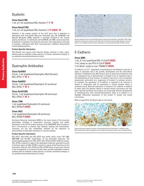
Human skeletal muscle: immunohistochemical staining for dystrophin using NCL-DYSA. Note<br />
membrane staining of normal muscle fibers (A) and reduced and variable staining of muscle<br />
fibers in an individual with Duchenne and Becker muscular dystrophy (B). Paraffin section.<br />
E-Cadherin<br />
Clone 36B5<br />
1 mL, 0.1 mL lyophilized NCL-E-Cad P (HIER)<br />
7 mL ready-to-use RTU-E-Cad P (HIER)<br />
7 mL Bond ready-to-use PA0387 P (HIER)<br />
E-cadherin is a Ca 2+ -dependent, transmembrane cell adhesion molecule. It<br />
plays an important role in the g<strong>row</strong>th, development and the intercellular<br />
adhesion of epithelial cells. Most tumors have an abnormal architecture and<br />
any subsequent loss of adhesiveness is thought to be an important step in<br />
the development of local invasion. E-cadherin may have a role in neoplastic<br />
progression, particularly as a suppressor of invasion. In prostate cancers,<br />
for example, the expression of E-cadherin is reported to be reduced or<br />
absent in comparison with its expression in normal prostate which is<br />
uniformly strong. Reduced expression or absence of E-cadherin in addition<br />
to alpha, beta and gamma-catenin in primary breast carcinomas has also<br />
been reported and these four proteins are associated with the development<br />
of metastases.The main immunohistochemical utility of E-cadherin is to<br />
highlight differential expression of this protein in lobular and ductal<br />
carcinomas.<br />
Refer to page 28 for the Bond ready-to-use format.<br />
Human pulmonary adenocarcinoma: immunohistochemical staining for E-cadherin using<br />
NCL-E-Cad. Note intense cytoplasmic and membrane staining of tumor cells. Paraffin section.<br />
Products in this catalog are subject to regulatory approval.<br />
This catalog is not for use in the USA.


