

Labelling Review row-Online
Labelling Review row-Online
Labelling Review row-Online

You also want an ePaper? Increase the reach of your titles
YUMPU automatically turns print PDFs into web optimized ePapers that Google loves.
Primary Antibodies<br />
LFA-2 (CD2)<br />
Clone AB75<br />
1 mL, 0.1 mL lyophilized NCL-CD2-271 P (HIER)<br />
1 mL liquid NCL-L-CD2-271 P (HIER)<br />
7 mL ready-to-use RTU-CD2-271 P (HIER)<br />
See also CD2 (LFA-2) on page 70.<br />
Linker for Activation of T Cells<br />
Clone 3.8<br />
1 mL liquid NCL-L-LAT F P (HIER)<br />
7 mL ready-to-use RTU-LAT F P (HIER)<br />
Stimulation of the T cell antigen receptor (TCR) results in the activation of<br />
several protein tyrosine kinases (PTKs) associated with the TCR. These<br />
activated PTKs phosphorylated tyrosine residues on multiple protein<br />
substrates. This phosphorylation results in the activation of enzymes such<br />
as phospholipase C gamma or creates sites of binding for proteins involved<br />
in the activation cascade. Linker for activation of T cells (LAT) is an integral<br />
membrane protein (36 to 38 kD) which plays an important role in linking<br />
engagement of the TCR to the biochemical events of T cell activation. LAT is<br />
a substrate of activated ZAP-70 and Syk PTKs. It binds following tyrosine<br />
phosphorylation, Grb2, PLC-gamma1 and other critical signalling molecules<br />
recruiting them to the plasma membrane. This has the effect of enhancing<br />
the phosphorylation of tyrosine residues required for enzymatic activation<br />
and promoting the formation of protein complexes. LAT mRNA is found in NK<br />
cells and mast cells. LAT protein has been reported to be detected in thymus<br />
and peripheral lymphoid tissues such as T cell areas in lymph nodes and<br />
spleen. In the small intestine, intra-epithelial T cells also express LAT, and in<br />
bone mar<strong>row</strong>, LAT is expressed by T lymphocytes in interstitial spaces and<br />
also by platelets and megakaryocytes. LAT is reported not to be expressed<br />
in B cells, macrophages, plasma cells, monocytes, epithelial histiocytes and<br />
dendritic cells.<br />
Human tonsil: immunohistochemical staining for LAT protein using NCL-L-LAT. Note intense<br />
membrane staining of T lymphocytes. Paraffin section.<br />
LMP-1 (Epstein-Barr virus)<br />
CS1, CS2, CS3, Clone CS4<br />
1 mL, 0.1 mL lyophilized NCL-EBV-CS1-4 F P (Enzyme)<br />
See also Epstein-Barr virus (LMP-1) on page 106.<br />
/ 128<br />
For detailed information on all products please visit our website:<br />
www.leica-microsystems.com<br />
L-selectin (CD62L)<br />
Clone 9H6<br />
1 mL lyophilized NCL-CD62L-489 P (HIER)<br />
The CD62L antigen is also known as Leu-8, TQ1, LAM1, MEL-14 antigen,<br />
lymph node homing antigen and L-selectin. It mediates the tethering and<br />
rolling of leukocytes on endothelial surfaces and also contributes to the<br />
recruitment of leukocytes from the blood to areas of inflammation. CD62L<br />
antigen is also important for the homing of naive lymphocytes to peripheral<br />
lymph nodes and Peyer's patches and can also mediate neutrophil to<br />
neutrophil interactions via the recognition of CD162 antigen. CD62L antigen<br />
is reported to be expressed on the surface of mantle zone B lymphocytes in<br />
different lymphoid sites but is absent on germinal center B cells. It is also<br />
expressed on a proportion of T cells in peripheral lymph nodes, mucosal<br />
lymphoid sites and spleen. Non-lymphocytic staining has been reported on<br />
Langerhans cells, follicular dendritic cells in tonsil, neutrophils, monocytes<br />
and on macrophages in the thymus. Ligands for CD62L are expressed not<br />
only in specific vascular endothelium in lymph nodes and Peyer's patches<br />
but also in extravascular tissues such as brain white matter, the choroid<br />
plexus and in kidney distal tubuli.<br />
Human tonsil: immunohistochemical staining for CD62L antigen (L-selectin) using<br />
NCL-CD62L-489. Note membrane staining of lymphocytes. Paraffin section.<br />
Lung Resistance-Related Protein (110 kD)<br />
Clone 9D6<br />
1 mL lyophilized NCL-LRRP P (HIER) W<br />
Multidrug-resistant cancer cell lines are reported to frequently overexpress<br />
the 100 kD lung resistance protein (LRP) also known as lung resistancerelated<br />
protein (LRRP). The overexpression of LRRP in acute myeloid<br />
leukemias, multiple and ovarian carcinomas has been reported. LRRP<br />
functions as a major vault transporter protein where vaults are multi-subunit<br />
structures which may be involved in nucleocytoplasmic transport. LRRP is<br />
overexpressed in P-glycoprotein negative multidrug-resistant tumor cell<br />
lines of different histogenetic origins and show an ATP-dependent drug<br />
accumulation effect. LRRP is also reported to be expressed in normal<br />
tissues, with expression being highest in epithelial cells with secretory and<br />
excretory functions such as bronchial cells and intestinal epithelial cells.<br />
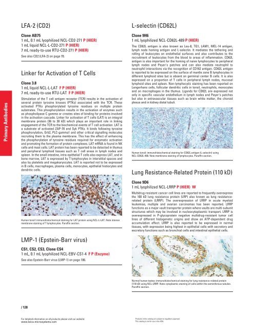
Normal human testes: immunohistochemical staining for lung resistance-related protein<br />
(110 kD) using NCL-LRRP. Note cytoplasmic staining of cells within the seminiferous tubules.<br />
Paraffin section.<br />
Products in this catalog are subject to regulatory approval.<br />
This catalog is not for use in the USA.


