

Labelling Review row-Online
Labelling Review row-Online
Labelling Review row-Online

Create successful ePaper yourself
Turn your PDF publications into a flip-book with our unique Google optimized e-Paper software.
Primary Antibodies<br />
CD1a<br />
Clone MTB1<br />
1 mL, 0.1 mL lyophilized NCL-CD1a-235 F P (HIER)<br />
1 mL liquid NCL-L-CD1a-235 F P (HIER)<br />
7 mL ready-to-use RTU-CD1a-235 F P (HIER)<br />
7 mL Bond ready-to-use PA0235 P (HIER)<br />
Clone JPM30<br />
1 mL, 0.1 mL lyophilized NCL-CD1a-220 F P (HIER)<br />
CD1a is a protein of 43 to 49 kD expressed on dendritic cells and cortical<br />
thymocytes. CD1a antigen expression has been shown to be useful in<br />
differentiating Langerhans cells, powerful antigen presenting cells present<br />
in skin and epithelia, from interdigitating cells. Immunohistochemical studies<br />
for CD1a antigen have reported a reduction in epidermal Langerhans cells in<br />
graft versus host disease and the participation of CD1a antigen-positive<br />
dendritic cells in atherosclerotic lesion formation and asthmatic<br />
inflammation.<br />
Product Specific Information<br />
Clone MTB1 detects cortical thymocytes, Langerhans cells in epidermis,<br />
interdigitating cells of dermis and interdigitating cells of stratified squamous<br />
epithelium of tonsil. Clone MTB1 may also detect small focal groups of<br />
lymphocytes outside the germinal centers of tonsil indicating a crossreaction<br />
with CD1b antigen. Clone JPM30 detects cortical thymocytes,<br />
Langerhans cells in epidermis, interdigitating cells of dermis, interdigitating<br />
cells of stratified squamous epithelium of tonsil but in addition it stains sweat<br />
gland ducts in the dermis and epithelial cells of small intestine indicative of<br />
cross-reactivity with CD1d antigen.<br />
Refer to page 17 for the Bond ready-to-use format.<br />
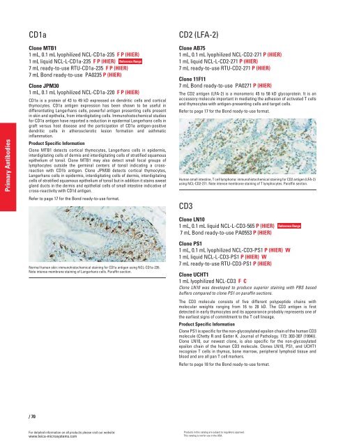
Normal human skin: immunohistochemical staining for CD1a antigen using NCL-CD1a-235.<br />
Note intense membrane staining of Langerhans cells. Paraffin section.<br />
/70<br />
For detailed information on all products please visit our website:<br />
www.leica-microsystems.com<br />
Reference Range<br />
CD2 (LFA-2)<br />
Clone AB75<br />
1 mL, 0.1 mL lyophilized NCL-CD2-271 P (HIER)<br />
1 mL liquid NCL-L-CD2-271 P (HIER)<br />
7 mL ready-to-use RTU-CD2-271 P (HIER)<br />
Clone 11F11<br />
7 mL Bond ready-to-use PA0271 P (HIER)<br />
The CD2 antigen (LFA-2) is a monomeric 45 to 58 kD glycoprotein. It is an<br />
accessory molecule important in mediating the adhesion of activated T cells<br />
and thymocytes with antigen-presenting cells and target cells.<br />
Refer to page 17 for the Bond ready-to-use format.<br />
Human small intestine, T cell lymphoma: immunohistochemical staining for CD2 antigen (LFA-2)<br />
using NCL-CD2-271. Note intense membrane staining of T lymphocytes. Paraffin section.<br />
CD3<br />
Clone LN10<br />
1 mL, 0.1 mL liquid NCL-L-CD3-565 P (HIER)<br />
7 mL Bond ready-to-use PA0553 P (HIER)<br />
Clone PS1<br />
1 mL, 0.1 mL lyophilized NCL-CD3-PS1 P (HIER) W<br />
1 mL liquid NCL-L-CD3-PS1 P (HIER) W<br />
7 mL ready-to-use RTU-CD3-PS1 P (HIER)<br />
Clone UCHT1<br />
1 mL lyophilized NCL-CD3 FC<br />
Clone LN10 was developed to produce superior staining with PBS based<br />
buffers compared to clone PS1 on paraffin sections.<br />
The CD3 molecule consists of five different polypeptide chains with<br />
molecular weights ranging from 16 to 28 kD. The CD3 antigen is first<br />
detected in early thymocytes and its appearance probably represents one of<br />
the earliest signs of commitment to the T cell lineage.<br />
Product Specific Information<br />
Clone PS1 is specific for the non-glycosylated epsilon chain of the human CD3<br />
molecule (Chetty R and Gatter K. Journal of Pathology. 173: 303-307 (1994)).<br />
Clone LN10, our newest clone, is also specific for the non-glycosylated<br />
epsilon chain of the human CD3 molecule. Clones LN10, PS1, and UCHT1<br />
recognize T cells in thymus, bone mar<strong>row</strong>, peripheral lymphoid tissue and<br />
blood and are all pan T cell markers.<br />
Refer to page 18 for the Bond ready-to-use format.<br />
Products in this catalog are subject to regulatory approval.<br />
This catalog is not for use in the USA.<br />
Reference Range


