

Labelling Review row-Online
Labelling Review row-Online
Labelling Review row-Online

You also want an ePaper? Increase the reach of your titles
YUMPU automatically turns print PDFs into web optimized ePapers that Google loves.
CD40<br />
Clone 11E9<br />
1 mL, 0.1 mL lyophilized NCL-CD40 P (HIER) W<br />
The CD40 antigen is a single chain glycoprotein with a calculated molecular<br />
weight of 27 kD. It is known to be a member of the tumor necrosis factor/<br />
nerve g<strong>row</strong>th factor superfamily and shows a significant homology to the<br />
Hodgkin's disease-associated antigen, CD30. The precise function of the<br />
CD40 antigen is unknown but it appears to be involved in the transduction of<br />
regulatory signals for cellular functions such as B cell proliferation and<br />
differentiation. It is also important in the prevention of apoptosis of germinal<br />
center B cells. The CD40 antigen is reported to be found on mature B cells<br />
(except plasma cells), most B cell leukemias and lymphomas, interdigitating<br />
reticulum cells, follicular dendritic cells and Reed Sternberg cells. Outside<br />
the immune system, CD40 antigen is reported to be expressed on some<br />
epithelial cells of certain carcinomas and in malignant melanomas.<br />
CD41 (GPIIb/IIIa)<br />
Clone M148<br />
1 mL lyophilized NCL-CD41 F<br />
The CD41 antigen, also known as GPIIb/IIIa, is reported to be expressed<br />
early in megakaryocyte maturation and in megakaryoblastic leukemias and<br />
is absent or defective in platelets from patients with Glanzmann's thrombasthenia.<br />
The CD41 antigen is involved in fibrinogen binding, clot retraction<br />
and platelet aggregation.<br />
Human tonsil: immunohistochemical staining for CD41 antigen (GPIIb/IIIa) using NCL-CD41.<br />
Note staining of aggregated platelets within the blood vessel. Frozen section.<br />
CD42b (GPIb)<br />
Clone MM2/174<br />
1 mL lyophilized NCL-CD42b F P (HIER)<br />
The CD42b glycoprotein, also known as GPIb, is a co-factor of ristocetininduced<br />
aggregation and is involved in the binding of platelets to blood<br />
vessel walls. The CD42b antigen is reported to be expressed on platelets and<br />
on megakaryocytes in bone mar<strong>row</strong> and in megakaryoblastic leukemias. The<br />
absence of CD42b antigen on platelets is reported to be a possible indicator<br />
of Bernard-Soulier disease.<br />
CD43<br />
Clone MT1<br />
1 mL lyophilized NCL-MT1 F P (Enzyme) W<br />
1 mL liquid NCL-L-MT1 F P (Enzyme) W<br />
7 mL ready-to-use RTU-MT1 F P (Enzyme)<br />
7 mL Bond ready-to-use PA0938 P (HIER)<br />
Clone MT1 produces superior staining on paraffin sections.<br />
The CD43 antigen is expressed on the membrane and in the cytoplasm of T<br />
cells and cells of myeloid lineage. The molecule itself exhibits molecular<br />
weight heterogeneity with bands of 90 to 140 kD observed on SDS-PAGE<br />
between different cell lines. Cells expressing the CD43 antigen are reported<br />
to include normal and neoplastic T cells. A small proportion of B cell chronic<br />
leukemias and centrocytic lymphomas are also reported to express CD43<br />
antigen.<br />
Product Specific Information<br />
Enzyme pretreatment may enhance staining with clone MT1 in some cases.<br />
Refer to page 22 for the Bond ready-to-use format.<br />
Human mantle cell lymphoma: immunohistochemical staining for CD43 antigen using NCL-MT1.<br />
Note intense membrane staining of tumor cells. Paraffin section.<br />
CD44 (H-CAM)<br />
Clone DF1485<br />
1 mL, 0.1 mL lyophilized NCL-CD44-2 F P (HIER) C<br />
The CD44 antigen (H-CAM) is an 80 to 95 kD transmembrane glycoprotein<br />
with extensive O-linked glycosylation. The antigen is a cell surface receptor<br />
for hyaluronate, suggesting a role in the regulation of cell substrate<br />
interactions as well as cell migration. CD44 antigen is reported to be<br />
expressed on T cells, B cells, monocytes, granulocytes, erythrocytes and<br />
weakly on platelets. Other CD44 antigen positive cell types are reported to<br />
include epithelial cells, glial cells, fibroblasts and myocytes. Increased<br />
expression of CD44 antigen is found on some carcinomas and it has been<br />
reported that transition of tumor cell lines from non-metastatic to metastatic<br />
may be associated with changes in the expression of CD44 antigen variants.<br />
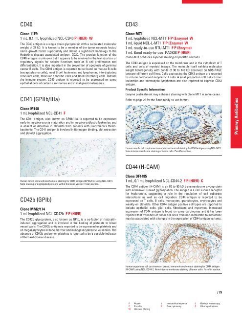
Human squamous cell carcinoma of breast: immunohistochemical staining for CD44 antigen<br />
(H-CAM) using NCL-CD44-2. Note intense membrane staining of tumor cells. Paraffin section.<br />
F Frozen I Immunofluorescence E Electron microscopy<br />
P Paraffin C Flow cytometry O Other applications<br />
W Western blotting<br />
/79<br />
Primary Antibodies


