

Labelling Review row-Online
Labelling Review row-Online
Labelling Review row-Online

Create successful ePaper yourself
Turn your PDF publications into a flip-book with our unique Google optimized e-Paper software.
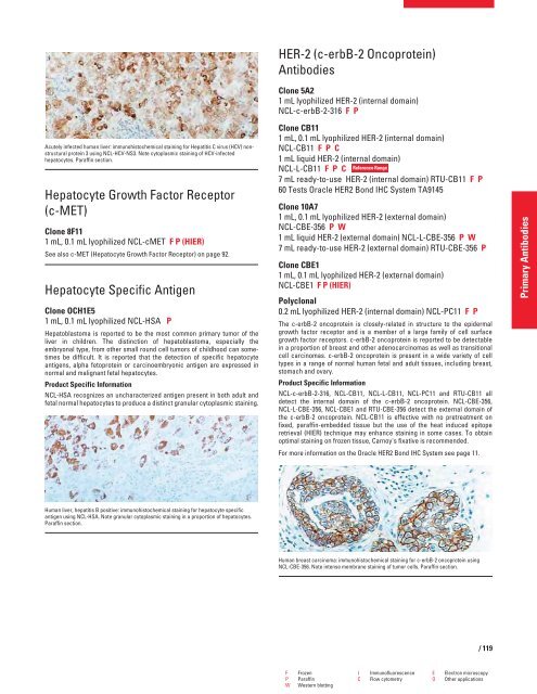
Acutely infected human liver: immunohistochemical staining for Hepatitis C virus (HCV) nonstructural<br />
protein 3 using NCL-HCV-NS3. Note cytoplasmic staining of HCV-infected<br />
hepatocytes. Paraffin section.<br />
Hepatocyte G<strong>row</strong>th Factor Receptor<br />
(c-MET)<br />
Clone 8F11<br />
1 mL, 0.1 mL lyophilized NCL-cMET F P (HIER)<br />
See also c-MET (Hepatocyte G<strong>row</strong>th Factor Receptor) on page 92.<br />
Hepatocyte Specific Antigen<br />
Clone OCH1E5<br />
1 mL, 0.1 mL lyophilized NCL-HSA P<br />
Hepatoblastoma is reported to be the most common primary tumor of the<br />
liver in children. The distinction of hepatoblastoma, especially the<br />
embryonal type, from other small round cell tumors of childhood can sometimes<br />
be difficult. It is reported that the detection of specific hepatocyte<br />
antigens, alpha fetoprotein or carcinoembryonic antigen are expressed in<br />
normal and malignant fetal hepatocytes.<br />
Product Specific Information<br />
NCL-HSA recognizes an uncharacterized antigen present in both adult and<br />
fetal normal hepatocytes to produce a distinct granular cytoplasmic staining.<br />
Human liver, hepatitis B positive: immunohistochemical staining for hepatocyte specific<br />
antigen using NCL-HSA. Note granular cytoplasmic staining in a proportion of hepatocytes.<br />
Paraffin section.<br />
HER-2 (c-erbB-2 Oncoprotein)<br />
Antibodies<br />
Clone 5A2<br />
1 mL lyophilized HER-2 (internal domain)<br />
NCL-c-erbB-2-316 FP<br />
Clone CB11<br />
1 mL, 0.1 mL lyophilized HER-2 (internal domain)<br />
NCL-CB11 FPC<br />
1 mL liquid HER-2 (internal domain)<br />
NCL-L-CB11 FPC<br />
Reference Range<br />
7 mL ready-to-use HER-2 (internal domain) RTU-CB11 FP<br />
60 Tests Oracle HER2 Bond IHC System TA9145<br />
Clone 10A7<br />
1 mL, 0.1 mL lyophilized HER-2 (external domain)<br />
NCL-CBE-356 PW<br />
1 mL liquid HER-2 (external domain) NCL-L-CBE-356 PW<br />
7 mL ready-to-use HER-2 (external domain) RTU-CBE-356 P<br />
Clone CBE1<br />
1 mL, 0.1 mL lyophilized HER-2 (external domain)<br />
NCL-CBE1 F P (HIER)<br />
Polyclonal<br />
0.2 mL lyophilized HER-2 (internal domain) NCL-PC11 FP<br />
The c-erbB-2 oncoprotein is closely-related in structure to the epidermal<br />
g<strong>row</strong>th factor receptor and is a member of a large family of cell surface<br />
g<strong>row</strong>th factor receptors. c-erbB-2 oncoprotein is reported to be detectable<br />
in a proportion of breast and other adenocarcinomas as well as transitional<br />
cell carcinomas. c-erbB-2 oncoprotein is present in a wide variety of cell<br />
types in a range of normal human fetal and adult tissues, including breast,<br />
stomach and ovary.<br />
Product Specific Information<br />
NCL-c-erbB-2-316, NCL-CB11, NCL-L-CB11, NCL-PC11 and RTU-CB11 all<br />
detect the internal domain of the c-erbB-2 oncoprotein. NCL-CBE-356,<br />
NCL-L-CBE-356, NCL-CBE1 and RTU-CBE-356 detect the external domain of<br />
the c-erbB-2 oncoprotein. NCL-CB11 is effective with no pretreatment on<br />
fixed, paraffin-embedded tissue but the use of the heat induced epitope<br />
retrieval (HIER) technique may enhance staining in some cases. To obtain<br />
optimal staining on frozen tissue, Carnoy's fixative is recommended.<br />
For more information on the Oracle HER2 Bond IHC System see page 11.<br />
Human breast carcinoma: immunohistochemical staining for c-erbB-2 oncoprotein using<br />
NCL-CBE-356. Note intense membrane staining of tumor cells. Paraffin section.<br />
F Frozen I Immunofluorescence E Electron microscopy<br />
P Paraffin C Flow cytometry O Other applications<br />
W Western blotting<br />
/ 119<br />
Primary Antibodies


