

Labelling Review row-Online
Labelling Review row-Online
Labelling Review row-Online

Create successful ePaper yourself
Turn your PDF publications into a flip-book with our unique Google optimized e-Paper software.
Primary Antibodies<br />
Epstein-Barr virus-Induced Gene 3<br />
Protein<br />
Clone EL8<br />
1 mL lyophilized NCL-EBI-3 F P (HIER)<br />
Epstein-Barr virus (EBV)-associated Hodgkin's lymphoma (HL) and nasopharyngeal<br />
carcinoma (NPC) usually occurs in individuals without clinically<br />
apparent deficiencies in anti-viral immunity. Despite expressing viral<br />
proteins, both tumors are apparently able to escape EBV-specific immunity<br />
in vivo. EBI-3 is an EBV-induced cytokine homologous to the interleukin 12<br />
p40 subunit which can heterodimerize with the interleukin 12 p35 subunit.<br />
Researchers have suggested that EBI-3 protein may function to antagonize<br />
interleukin 12 and to inhibit the development of a Th1 immune response. It<br />
has been reported that EBI-3 protein is strongly expressed in Hodgkin's<br />
Reed Sternberg (RS) cells in approximately 96 percent of HL cases,<br />
independently of the EBV status of the tumor cells. EBI-3 protein has also<br />
been reported to be detected in a small percentage of epithelial tumor cells<br />
of NPC biopsies but not in Burkitt's lymphomas. EBI-3 protein may be an<br />
additional component of the repertoire employed by Hodgkin's RS cells to<br />
inhibit and effect anti-tumor or anti-viral immune response. EBI-3 protein<br />
expression has also been reported in spleen, tonsil, mature dendritic cells,<br />
colonic mucosa and at high levels in full term placenta.<br />
Hodgkin's lymphoma: immunohistochemical staining for Epstein-Barr virus-induced gene 3<br />
protein using NCL-EBI-3. Note cytoplasmic staining of infected cells. Paraffin section.<br />
Epstein-Barr virus (LMP-1)<br />
Clone CS1/CS2/CS3/CS4 cocktail<br />
1 mL, 0.1 mL lyophilized NCL-EBV-CS1-4 F P (Enzyme)<br />
Epstein-Barr virus (EBV) is one of the eight known human herpes viruses<br />
and belongs to the Gammaherpes viriniae, the same subfamily as human<br />
herpesvirus type 8 (HHV-8). Herpes viruses have large double strand DNA<br />
genomes and are complex viruses often encoding over 35 proteins including<br />
enzymes involved in nucleic acid metabolism, DNA synthesis and protein<br />
processing in addition to viral structural proteins. These viruses are capable<br />
of entering a latent phase where the host shows no visible signs of infection<br />
and levels of infectious agent become very low. During latency, viral gene<br />
expression is restricted to only a few genes. Latent membrane protein (LMP-<br />
1) is a 60 kD protein encoded by the BNLF1 gene of EBV.<br />
Product Specific Information<br />
NCL-EBV-CS1-4 is a cocktail of four monoclonal antibodies; clones CS1, CS2,<br />
CS3 and CS4.<br />
/ 106<br />
For detailed information on all products please visit our website:<br />
www.leica-microsystems.com<br />
Epstein-Barr virus (nuclear antigen 2)<br />
Clone PE2<br />
1 mL, 0.1 mL lyophilized NCL-EBV-PE2 FW<br />
Epstein-Barr virus (EBV) nuclear antigen 2 (EBNA2) is an EBV-encoded<br />
nuclear protein of 82 kD. EBNA2 is essential for g<strong>row</strong>th transformation of<br />
B lymphocytes and has been shown to modulate the activity of several viral<br />
and cellular promoters.<br />
Estrogen Receptor<br />
Clone 6F11<br />
2 mL lyophilized NCL-ER-6F11/2 F P (HIER) W C<br />
2 mL liquid NCL-L-ER-6F11/2 F P (HIER) W C<br />
1 mL. 0.1mL lyophilized NCL-ER-6F11 F P (HIER) W C<br />
1 mL liquid NCL-L-ER-6F11 F P (HIER) W C<br />
7 mL ready-to-use RTU-ER-6F11 F P (HIER) W<br />
7 mL Bond ready-to-use PA0151 P (HIER)<br />
Estrogen receptor (ER) content of breast cancer tissue is an important<br />
parameter in the prediction of prognosis and response to endocrine therapy.<br />
The introduction of highly specific monoclonal antibodies to ER has allowed<br />
the determination of receptor status of breast tumors to be carried out in<br />
routine histopathology laboratories.<br />
Product Specific Information<br />
Clone 6F11 is raised to the full length alpha form of the estrogen receptor<br />
molecule present on human ER antigen, located in the nucleus of ER positive<br />
normal and neoplastic cells. Clone 6F11 has been extensively tested (Bevitt D<br />
J et al. Journal of Pathology. 183 : 228-232 (1997)). Further publications exist<br />
that discuss the sensitivity of clone 6F11 (Kauffman O et al. Modern Pathology<br />
11(4):357-363 (1998)) and Kaplan P A et al. American Journal of Clinical<br />
Pathology 123: 276-280 (2005). NCL-ER-6F11/2 and NCL-L-ER-6F11/2 are more<br />
economic options for high volume users of clone 6F11.<br />
Refer to page 28 for the Bond ready-to-use format.<br />
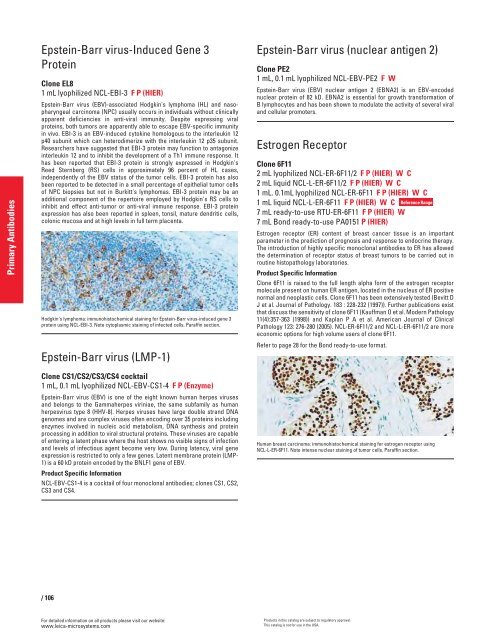
Human breast carcinoma: immunohistochemical staining for estrogen receptor using<br />
NCL-L-ER-6F11. Note intense nuclear staining of tumor cells. Paraffin section.<br />
Products in this catalog are subject to regulatory approval.<br />
This catalog is not for use in the USA.<br />
Reference Range


