

Labelling Review row-Online
Labelling Review row-Online
Labelling Review row-Online

Create successful ePaper yourself
Turn your PDF publications into a flip-book with our unique Google optimized e-Paper software.
Product Specific Information<br />
Using retrieval solutions other than that recommended for Clone JCM182 in<br />
the datasheet may increase background reactivity.<br />
Refer to page 21 for the Bond ready-to-use format.<br />
Hodgkin's lymphoma: immunohistochemical staining for CD30 antigen using NCL-L-CD30-591.<br />
Note membrane staining and characteristic staining of paranuclear hofs of Reed Sternberg<br />
cells. Paraffin section.<br />
CD31 (PECAM-1)<br />
Clone 1A10<br />
1 mL, 0.1 mL lyophilized NCL-CD31-1A10 P (HIER)<br />
7 mL Bond ready-to-use PA0250 P (HIER)<br />
CD31 antigen (PECAM-1) is a single chain transmembrane glycoprotein with<br />
a molecular weight of 130 to 140 kD. The CD31 molecule is expressed on the<br />
surface of platelets, monocytes, granulocytes, B cells and at the endothelial<br />
intracellular junction. The molecule has an extracellular domain that<br />
contains six Ig-like homology units of C2 subclass, typical of cell to cell<br />
adhesion molecules. This domain mediates endothelial cell to cell adhesion,<br />
plays a role in endothelial contact and may serve to stabilize the endothelial<br />
cell monolayer. The CD31 molecule also has a cytoplasmic domain with<br />
potential sites for phosphorylation after cellular activation. The properties of<br />
CD31 antigen suggest that it is involved in interactive events during<br />
angiogenesis, thrombosis and wound healing. Angiogenesis is essential for<br />
tumor g<strong>row</strong>th and metastases.<br />
Refer to page 21 for the Bond ready-to-use format.<br />
Human glomangioma: immunohistochemical staining for CD31 antigen (PECAM-1) using<br />
NCL-CD31-1A10. Note intense membrane staining of endothelial cells. Paraffin section.<br />
CD33<br />
Clone PWS44<br />
1 mL, 0.1 mL liquid NCL-L-CD33 P (HIER) W<br />
7 mL Bond ready-to-use PA0555 P (HIER)<br />
CD33 antigen is reported to appear on myelomonocytic precursor cells after<br />
CD34 antigen expression. It then continues to be expressed on both the<br />
myeloid and monocyte lineages, although it is reported to be absent on<br />
granulocytes. It has been reported that expression of CD33 is restricted to<br />
monocytes, premyelocytes, myeloid blasts, some acute undifferentiated<br />
leukemias and acute lymphoblastic leukemias. The expression of CD33<br />
antigen has been demonstrated to be an important marker for distinguishing<br />
myeloid from the lymphoid leukemias.<br />
Refer to page 22 for the Bond ready-to-use format.<br />
Acute myeloid leukemia: immunohistochemical staining for CD33 antigen using NCL-L-CD33.<br />
Note intense cytoplasmic and membrane staining of malignant cells. Paraffin section.<br />
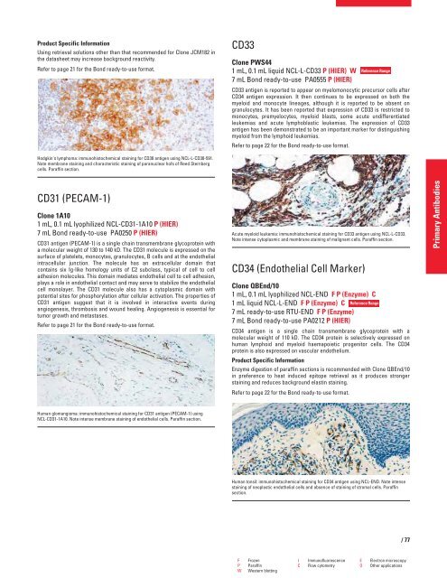
CD34 (Endothelial Cell Marker)<br />
Reference Range<br />
Clone QBEnd/10<br />
1 mL, 0.1 mL lyophilized NCL-END F P (Enzyme) C<br />
1 mL liquid NCL-L-END F P (Enzyme) C<br />
7 mL ready-to-use RTU-END F P (Enzyme)<br />
7 mL Bond ready-to-use PA0212 P (HIER)<br />
Reference Range<br />
CD34 antigen is a single chain transmembrane glycoprotein with a<br />
molecular weight of 110 kD. The CD34 protein is selectively expressed on<br />
human lymphoid and myeloid haemapoietic progenitor cells. The CD34<br />
protein is also expressed on vascular endothelium.<br />
Product Specific Information<br />
Enzyme digestion of paraffin sections is recommended with Clone QBEnd/10<br />
in preference to heat induced epitope retrieval as it produces stronger<br />
staining and reduces background elastin staining.<br />
Refer to page 22 for the Bond ready-to-use format.<br />
Human tonsil: immunohistochemical staining for CD34 antigen using NCL-END. Note intense<br />
staining of neoplastic endothelial cells and absence of staining of stromal cells. Paraffin<br />
section.<br />
F Frozen I Immunofluorescence E Electron microscopy<br />
P Paraffin C Flow cytometry O Other applications<br />
W Western blotting<br />
/77<br />
Primary Antibodies


