

Labelling Review row-Online
Labelling Review row-Online
Labelling Review row-Online

Create successful ePaper yourself
Turn your PDF publications into a flip-book with our unique Google optimized e-Paper software.
CD45RB<br />
Clone MEM55<br />
1 mL lyophilized NCL-CD45RB F P (HIER)<br />
The CD45 molecule is reported to be found on all cells of hematopoietic<br />
origin, except erythrocytes. The various isoforms are expressed differently<br />
on various lymphoid cell types and are termed CD45RA, CD45RB, CD45RC<br />
and CD45RO. Low expression of CD45RB on CD45RO positive T lymphocytes<br />
defines a subset of highly differentiated T lymphocytes which accumulate in<br />
vivo within affected rheumatoid arthritic joints. The percentage of these cell<br />
types is also reported to be increased in the circulation of individuals with<br />
acute EBV infection and it is thought that these cells have a migratory<br />
advantage and are selectively recruited to sites of inflammation. CD45RB<br />
antigen is also reported to be found on B cells, monocytes, macrophages<br />
and is expressed weakly on granulocytes.<br />
Product Specific Information<br />
The heat induced epitope retrieval (HIER) technique may enhance staining in<br />
some cases.<br />
Human stomach, B cell lymphoma: immunohistochemical staining for CD45RB antigen using<br />
NCL-CD45RB. Note intense membrane staining of neoplastic lymphoid cells. Paraffin section.<br />
CD45RO<br />
Clone UCHL1<br />
1 mL, 0.1 mL lyophilized NCL-UCHL1 F P (HIER) C<br />
1 mL liquid NCL-L-UCHL1 F P (HIER) C<br />
7 mL ready-to-use RTU-UCHL1 F P (HIER)<br />
7 mL Bond ready-to-use PA0146 P (HIER)<br />
The CD45RO molecule, a 180 kD isoform of CD45, is reported to be expressed<br />
on 48 percent of peripheral blood T lymphocytes, 37 percent of CD4 positive<br />
lymphocytes, 80 percent of thymocytes and on the majority of T cell<br />
malignancies. Monocytes and granulocytes show surface expression of the<br />
antigen whereas tissue macrophages exhibit cytoplasmic expression. The<br />
heat induced epitope retrieval (HIER) technique may enhance staining in<br />
some cases.<br />
Refer to page 23 for the Bond ready-to-use format.<br />
CD54 (ICAM-1)<br />
Clone 23G12<br />
1 mL, 0.1 mL lyophilized NCL-CD54-307 P (HIER)<br />
See also ICAM-1 (CD54) on page 123.<br />
CD56 (NCAM)<br />
Clone CD564<br />
1 mL, 0.1 mL lyophilized NCL-CD56-504 P (HIER)<br />
7 mL Bond ready-to-use PA0191 P (HIER)<br />
Clone 1B6<br />
1 mL, 0.1 mL lyophilized NCL-CD56-1B6 P (HIER) W<br />
1 mL liquid NCL-L-CD56-1B6 P (HIER) W<br />
1 mL ready-to-use RTU-CD56-1B6 P (HIER)<br />
Clone CD564 was developed to produce superior staining on paraffin<br />
sections.<br />
The neural cell adhesion molecules are a family of closely-related cell<br />
surface glycoproteins thought to play a role in embryogenesis, development<br />
and contact-mediated interactions between neural cells. The CD56 antigen<br />
(NCAM) consists of four major isoforms generated by differential splicing of<br />
the RNA transcript from a single gene located on chromosome 5. The CD56<br />
antigen is expressed on neurons, astrocytes, Schwann cells, NK cells and a<br />
subset of activated T lymphocytes.<br />
Refer to page 23 for the Bond ready-to-use format.<br />
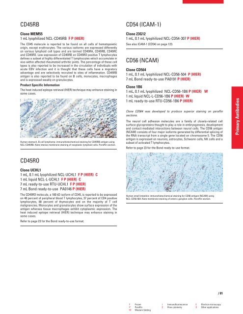
Human small intestine: immunohistochemical staining for CD56 antigen (NCAM) using<br />
NCL-CD56-564. Note membrane staining of enteric ganglion cells. Paraffin section.<br />
F Frozen I Immunofluorescence E Electron microscopy<br />
P Paraffin C Flow cytometry O Other applications<br />
W Western blotting<br />
/81<br />
Primary Antibodies


