

Labelling Review row-Online
Labelling Review row-Online
Labelling Review row-Online

Create successful ePaper yourself
Turn your PDF publications into a flip-book with our unique Google optimized e-Paper software.
Primary Antibodies<br />
CD7<br />
Clone LP15<br />
1 mL, 0.1 mL liquid NCL-L-CD7-580 P (HIER)<br />
7 mL Bond ready-to-use PA0266 P (HIER)<br />
The CD7 molecule is a membrane-bound glycoprotein of 40 kD and is the<br />
earliest T cell specific antigen to be expressed in lymphocytes. CD7 antigen<br />
is also the only early marker to persist throughout differentiation. The<br />
function and role of the CD7 molecule has not yet been fully identified,<br />
although the activation of T cells with gamma/delta receptors has been<br />
proposed based on mAb-induced activation. CD7 antigen is reported to be<br />
found on the majority of peripheral blood T cells, most natural killer cells and<br />
thymocytes.<br />
Product Specific Information<br />
Clone LP15 was developed to provide superior staining to clone CD7-272 on<br />
paraffin sections.<br />
Refer to page 18 for the Bond ready-to-use format.<br />
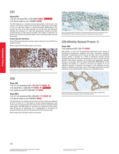
Human thymus: immunohistochemical staining for CD7 antigen using NCL-L-CD7-580.<br />
Note intense staining of cortical thymocytes. Paraffin section.<br />
CD8<br />
Clone 1A5<br />
1 mL, 0.1 mL lyophilized NCL-CD8-295 F P (HIER) W<br />
1 mL liquid NCL-L-CD8-295 F P (HIER) W<br />
7 mL ready-to-use RTU-CD8-295 F P (HIER)<br />
Clone 4B11<br />
1 mL, 0.1 mL lyophilized NCL-CD8-4B11 F P (HIER) W<br />
7 mL Bond ready-to-use PA0183 P (HIER)<br />
The CD8 molecule is composed of two chains and has a molecular weight of<br />
32 kD. It is found on a T cell subset of normal cytotoxic/suppressor cells<br />
which make up approximately 20 to 35 percent of human peripheral blood<br />
lymphocytes. The CD8 antigen is reported to be detected on natural killer<br />
cells, 80 percent of thymocytes, on a subpopulation of 30 percent of<br />
peripheral blood null cells and 15 to 30 percent of bone mar<strong>row</strong> cells.<br />
Refer to page 19 for the Bond ready-to-use format.<br />
/72<br />
For detailed information on all products please visit our website:<br />
www.leica-microsystems.com<br />
Reference Range<br />
Reference Range<br />
Large T cell immunoblastic lymphoma: immunohistochemical staining for CD8 antigen using<br />
NCL-CD8-295. Note intense membrane staining of T lymphocytes. Paraffin section.<br />
CD9 (Motility-Related Protein-1)<br />
Clone 72F6<br />
1 mL lyophilized NCL-CD9 F P (HIER)<br />
CD9 antigen is a 24 to 27 kD glycoprotein expressed on the surface of<br />
developing B lymphocytes, platelets, monocytes, eosinophils, basophils,<br />
stimulated T lymphocytes and by neurons and glial cells in the peripheral<br />
nervous system. It belongs to a family of membrane proteins termed<br />
tetraspanins which transverse the membrane four times. In pre-B cells and<br />
platelets, CD9 antigen regulates cell activation and aggregation possibly<br />
through an association with the integrin CD41/CD61 (GPIIb/GPIIIa). It also<br />
regulates cell motility in a variety of cell lines and appears to be an<br />
important regulator of Schwann cell behavior in the peripheral nervous<br />
system. In melanoma and breast cancer, CD9 antigen expression has been<br />
reported to occur predominantly on primary, non-metastatic tumors.<br />
Human fibroadenoma: immunohistochemical staining for CD9 antigen using NCL-CD9.<br />
Note intense membrane staining of tumor cells. Paraffin section.<br />
Products in this catalog are subject to regulatory approval.<br />
This catalog is not for use in the USA.


