Labelling Review row-Online
Labelling Review row-Online
Labelling Review row-Online
You also want an ePaper? Increase the reach of your titles
YUMPU automatically turns print PDFs into web optimized ePapers that Google loves.
Primary Antibodies<br />
Villin<br />
Clone CWWB1<br />
1 mL, 0.1 mL lyophilized NCL-VILLIN F P (HIER) W<br />
1 mL liquid NCL-L-VILLIN F P (HIER) W<br />
7 mL Bond ready-to-use PA0106 P (HIER)<br />
Villin and the structurally-related proteins gelsolin, fragmin and severin, all<br />
regulate the framework and assembly of actin. Villin is unique among these<br />
proteins in its ability to cross-link actin filaments into bundles, a process<br />
observed only at low Ca 2+ concentration. Villin is composed of three<br />
domains. The first two domains are homologous and the third domain is<br />
called the “headpiece”. This “headpiece” region is located at the C-terminus.<br />
Villin is mainly produced by epithelial cells that develop a brush border. Cells<br />
producing villin are reported to be found either in the epithelial cells of the<br />
intestinal mucosa and gall bladder, or in epithelial cells of the kidney<br />
proximal tubules and ductuli efferentes of the testis. However, villin is also<br />
reported to be found in some epithelia which lack a brush border but which<br />
are derived from embryonic gut such as duct cells of the exocrine pancreas<br />
and biliary cells of the liver. In these cell types, villin is concentrated in the<br />
apical cytoplasm. Epithelial cells of the intestinal mucosa are continually<br />
being renewed and this involves a migration of these cell types from the<br />
intestinal crypts to the tips of the villi, gradually acquiring their differentiated<br />
phenotype as they do so. The maximum production of villin occurs at the<br />
base of the villus. Villin, therefore, shows tissue-specific expression being<br />
restricted to certain epithelia and their apical domains, thus indicating their<br />
polarity. The morphological loss of polarity of colonic epithelial cells is<br />
reported to be one of the most significant indicators of dysplasia or<br />
neoplasia.<br />
Refer to page 43 for the Bond ready-to-use format.<br />
Vimentin<br />
Clone SRL33<br />
1 mL, 0.1 mL liquid NCL-L-VIM-572 P (HIER) W<br />
7 mL Bond ready-to-use PA0033 P (HIER)<br />
Clone V9<br />
1 mL, 0.1 mL lyophilized NCL-VIM-V9 F P (HIER) W<br />
1 mL liquid NCL-L-VIM-V9 F P (HIER) W<br />
7 mL ready-to-use RTU-VIM-V9 F P (HIER)<br />
Clone VIM 3B4<br />
1 mL lyophilized NCL-VIM F P (Enzyme) W<br />
Eukaryotic cells contain a number of types of cytoplasmic filamentous<br />
proteins, microtubule, microfilaments and intermediate-sized filaments (IF).<br />
Vimentin, a 57 kD protein that is an intermediate filament is reported to be<br />
expressed in most cells of mesenchymal origin, including fibroblasts,<br />
endothelial cells, smooth muscle, melanocytes as well as T and B<br />
lymphocytes.<br />
Refer to page 43 for the Bond ready-to-use format.<br />
/ 170<br />
For detailed information on all products please visit our website:<br />
www.leica-microsystems.com<br />
Reference Range<br />
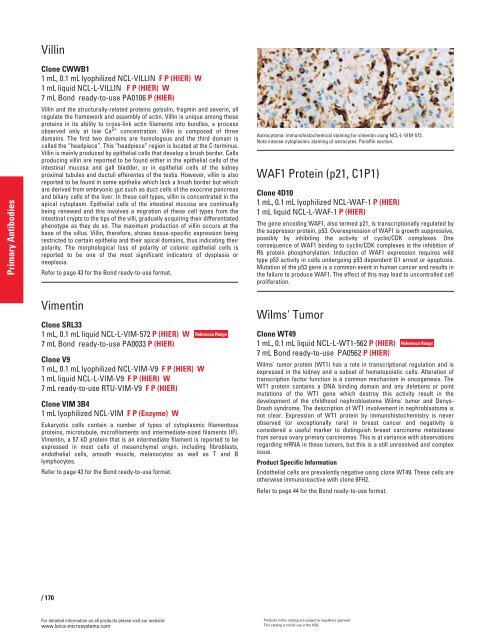
Astrocytoma: immunohistochemical staining for vimentin using NCL-L-VIM-572.<br />
Note intense cytoplasmic staining of astrocytes. Paraffin section.<br />
WAF1 Protein (p21, C1P1)<br />
Clone 4D10<br />
1 mL, 0.1 mL lyophilized NCL-WAF-1 P (HIER)<br />
1 mL liquid NCL-L-WAF-1 P (HIER)<br />
The gene encoding WAF1, also termed p21, is transcriptionally regulated by<br />
the suppressor protein, p53. Overexpression of WAF1 is g<strong>row</strong>th suppressive,<br />
possibly by inhibiting the activity of cyclin/CDK complexes. One<br />
consequence of WAF1 binding to cyclin/CDK complexes is the inhibition of<br />
Rb protein phosphorylation. Induction of WAF1 expression requires wild<br />
type p53 activity in cells undergoing p53 dependent G1 arrest or apoptosis.<br />
Mutation of the p53 gene is a common event in human cancer and results in<br />
the failure to produce WAF1. The effect of this may lead to uncontrolled cell<br />
proliferation.<br />
Wilms' Tumor<br />
Clone WT49<br />
1 mL, 0.1 mL liquid NCL-L-WT1-562 P (HIER)<br />
7 mL Bond ready-to-use PA0562 P (HIER)<br />
Wilms' tumor protein (WT1) has a role in transcriptional regulation and is<br />
expressed in the kidney and a subset of hematopoietic cells. Alteration of<br />
transcription factor function is a common mechanism in oncogenesis. The<br />
WT1 protein contains a DNA binding domain and any deletions or point<br />
mutations of the WT1 gene which destroy this activity result in the<br />
development of the childhood nephroblastoma Wilms' tumor and Denys-<br />
Drash syndrome. The description of WT1 involvement in nephroblastoma is<br />
not clear. Expression of WT1 protein by immunohistochemistry is never<br />
observed (or exceptionally rare) in breast cancer and negativity is<br />
considered a useful marker to distinguish breast carcinoma metastases<br />
from serous ovary primary carcinomas. This is at variance with observations<br />
regarding mRNA in these tumors, but this is a still unresolved and complex<br />
issue.<br />
Product Specific Information<br />
Endothelial cells are prevalently negative using clone WT49. These cells are<br />
otherwise immunoreactive with clone 6FH2.<br />
Refer to page 44 for the Bond ready-to-use format.<br />
Products in this catalog are subject to regulatory approval.<br />
This catalog is not for use in the USA.<br />
Reference Range


