

Labelling Review row-Online
Labelling Review row-Online
Labelling Review row-Online

Create successful ePaper yourself
Turn your PDF publications into a flip-book with our unique Google optimized e-Paper software.
p53 Protein<br />
Clone IMX25<br />
1 mL lyophilized NCL-p53-505 P (HIER) W<br />
7 mL Bond ready-to-use PA0057 P (HIER)<br />
p53 protein plays a vital role in suppressing the development of cancer. The<br />
accumulation of p53 protein in response to DNA damage in vitro is well<br />
established and appears to induce g<strong>row</strong>th arrest and apoptosis by the<br />
transcriptional regulation of other genes. In irradiated mice, p53 protein<br />
accumulates in splenocytes, thymocytes and osteocytes, though not in<br />
hepatocytes. Mouse T3T3 cells express high levels of phenotypically<br />
characteristic wild type p53 protein which carries two missense mutations.<br />
The range of antigenic sites in mouse p53 protein and human p53 protein is<br />
very similar.<br />
Product Specific Information<br />
NCL-p53-505 is raised to the same recombinant mouse p53 protein as the<br />
polyclonal, NCL-p53-CM5p. It reacts with rat and mouse p53 protein.<br />
Refer to page 38 for the Bond ready-to-use format.<br />
Mouse T3T3 cells: immunohistochemical staining for p53 mouse protein using NCL-p53-505.<br />
Note intense nuclear staining of a proportion of T3T3 cells. Paraffin section.<br />
p53 Protein (1801)<br />
Clone PAb 1801<br />
1 mL, 0.1 mL lyophilized NCL-p53-1801 F P (HIER) W C<br />
The gene for p53 is located on chromosome 17p, a frequent site of allelic<br />
loss in many tumors. It has been reported that a high proportion of breast<br />
and colon carcinomas show immunostaining for p53 protein and expression<br />
of p53 protein.<br />
Product Specific Information<br />
Clone PAb 1801 recognizes both wild type and mutant forms of human p53<br />
protein under denaturing and non-denaturing conditions.<br />
Human colonic adenocarcinoma: immunohistochemical staining for p53 protein using<br />
NCL-p53-1801. Note intense nuclear staining of tumor cells. Paraffin section.<br />
p53 Protein (BP53-12)<br />
Clone BP53-12<br />
1 mL lyophilized NCL-p53-BP FPW<br />
p53 protein plays a vital role in suppressing the development of cancer. The<br />
accumulation of p53 protein in response to DNA damage in vitro is well<br />
established and appears to induce g<strong>row</strong>th arrest and apoptosis by the<br />
transcriptional regulation of other genes.<br />
Product Specific Information<br />
Clone BP53-12 recognizes both wild type and mutant forms of human p53<br />
protein under denaturing and non-denaturing conditions. The heat induced<br />
epitope retrieval technique may improve staining in some cases.<br />
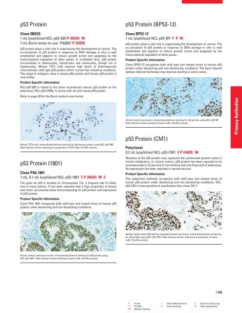
Human breast carcinoma: immunohistochemical staining for p53 protein using NCL-p53-BP.<br />
Note intense nuclear staining of tumor cells. Paraffin section.<br />
p53 Protein (CM1)<br />
Polyclonal<br />
0.2 mL lyophilized NCL-p53-CM1 F P (HIER) W<br />
Mutation of the p53 protein may represent the commonest genetic event in<br />
human malignancy. In colonic tumors, p53 protein has been reported to be<br />
overexpressed in 47 percent of carcinomas and only 9 percent of adenomas.<br />
No expression has been reported in normal mucosa.<br />
Product Specific Information<br />
This polyclonal antibody recognizes both wild type and mutant forms of<br />
human p53 protein under denaturing and non-denaturing conditions. NCLp53-CM1<br />
is less sensitive to overfixation than clone DO-7.<br />
Axillary lymph node infiltrated by metastatic breast carcinoma: immunohistochemical staining<br />
for p53 protein using NCL-p53-CM1. Note intense nuclear staining of a proportion of tumor<br />
cells. Paraffin section.<br />
F Frozen I Immunofluorescence E Electron microscopy<br />
P Paraffin C Flow cytometry O Other applications<br />
W Western blotting<br />
/ 145<br />
Primary Antibodies


