

Labelling Review row-Online
Labelling Review row-Online
Labelling Review row-Online

Create successful ePaper yourself
Turn your PDF publications into a flip-book with our unique Google optimized e-Paper software.
Bond<br />
CD68<br />
Clone 514H12<br />
7 mL Bond ready-to-use PA0273 P (HIER)<br />
For In Vitro Diagnostic Use<br />
The CD68 antigen is an intracellular molecule, which has primarily been<br />
associated with cytoplasmic granules and, to a lesser extent, the membranes<br />
of macrophages, monocytes, neutrophils, basophils and large<br />
lymphocytes. CD68 expression has been reported in stimulated T cells, NK<br />
cells, lymphomas, sarcomas and carcinomas, and in liver and renal tubules.<br />
Product Specific Information<br />
The CD68 antigen can be identified in a variety of normal and neoplastic<br />
tissues using CD68 (514H12).<br />
Also available as a Novocastra concentrate, refer to page 83.<br />
Tonsil: immunohistochemical staining with Bond ready-to-use CD68 (514H12) using Bond<br />
Polymer Refine Detection.<br />
CD79a<br />
Clone 11E3<br />
7 mL Bond ready-to-use PA0192 P (HIER)<br />
For In Vitro Diagnostic Use<br />
The CD79 complex is associated with membrane-bound immunoglobulins on<br />
B cells, with these immunoglobulins the two subunits, CD79a and CD79b<br />
constitute the B cell antigen receptor. The CD79a component is reported to<br />
first appear at the pre-B cell stage, early maturation, and persists until the<br />
plasma cell stage where it is found as an intracellular component. The<br />
CD79a antigen is reported to be expressed in the majority of acute leukemias<br />
of precursor B cell type, B cell lines, B cell lymphomas and in some<br />
myelomas. It is not present in myeloid or T cell lines.<br />
Also available as a Novocastra concentrate, refer to page 84.<br />
Tonsil, B cell-plasma cell transition: immunohistochemistry staining with Bond ready-to-use<br />
CD79a (11E3) using Bond Polymer Refine Detection.<br />
/24<br />
For detailed information on all products please visit our website:<br />
www.leica-microsystems.com<br />
CD99<br />
Clone 12E7<br />
7 mL Bond ready-to-use PA0509 P<br />
For In Vitro Diagnostic Use<br />
CD99 antigen, a 32 kD glycoprotein, is also known as MIC2, E2, 12E7, HuLym6<br />
or FMC29. CD99 antigen is reported to be expressed on cortical<br />
thymocytes and T lymphocytes and is involved in rosette formation with<br />
sheep or human erythrocytes. It is also expressed on granulosa cells of the<br />
ovary, most pancreatic islet cells, Sertoli cells of the testis and on some<br />
endothelial cells. CD99 antigen is reported to be strongly expressed on<br />
Ewing's sarcoma cells and primitive peripheral neuroectodermal tumors.<br />
Also available as a Novocastra concentrate, refer to page 85.<br />
Ewing’s sarcoma: immunohistochemical staining with Bond ready-to-use CD99 (12E7) using<br />
Bond Polymer Refine Detection.<br />
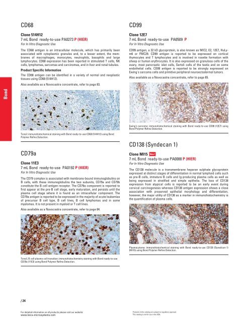
CD138 (Syndecan 1)<br />
Clone MI15 New!<br />
7 mL Bond ready-to-use PA0088 P (HIER)<br />
For In Vitro Diagnostic Use<br />
The CD138 molecule is a transmembrane heparan sulphate glycoprotein<br />
expressed at distinct stages of differentiation in normal lymphoid cells such<br />
as pre-B cells, immature B cells and Ig-producing plasma cells as well as<br />
being expressed in stratified and simple epithelia. The loss of CD138<br />
expression from atypical cells is reported to be an early event during<br />
cervical carcinogenesis whereas CD138 antigen expression shows a close<br />
association with preserved epithelial morphology and differentiation,<br />
however, the major utility of CD138 as a marker in immunohistochemistry is<br />
the quantification of plasma cells.<br />
Plasmacytoma: immunohistochemical staining with Bond ready-to-use CD138 (Synedcan-1)<br />
(MI15) using Bond Polymer Refine Detection.<br />
Products in this catalog are subject to regulatory approval.<br />
This catalog is not for use in the USA.


