

Labelling Review row-Online
Labelling Review row-Online
Labelling Review row-Online

Create successful ePaper yourself
Turn your PDF publications into a flip-book with our unique Google optimized e-Paper software.
Primary Antibodies<br />
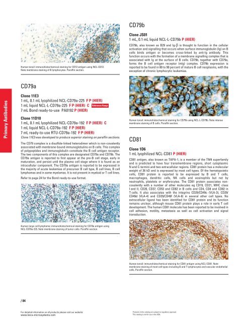
Human tonsil: immunohistochemical staining for CD72 antigen using NCL-CD72.<br />
Note membrane staining of B lymphocytes. Paraffin section.<br />
CD79a<br />
Clone 11E3<br />
1 mL, 0.1 mL lyophilized NCL-CD79a-225 F P (HIER)<br />
1 mL liquid NCL-L-CD79a-225 F P (HIER) C<br />
7 mL Bond ready-to-use PA0192 P (HIER)<br />
Clone 11D10<br />
1 mL, 0.1 mL lyophilized NCL-CD79a-192 F P (HIER) C<br />
1 mL liquid NCL-L-CD79a-192 F P (HIER)<br />
7 mL ready-to-use RTU-CD79a-192 F P (HIER)<br />
Clone 11E3 was developed to produce superior staining on paraffin sections.<br />
The CD79 complex is a disulfide-linked heterodimer which is non-covalently<br />
associated with membrane-bound immunoglobulins on B cells. This complex<br />
of polypeptides and immunoglobulin constitute the B cell antigen receptor.<br />
The two components of this complex are designated CD79a and CD79b. The<br />
CD79a antigen is reported to first appear at the pre-B cell stage, early in<br />
maturation, and persist until the plasma cell stage where it is found as an<br />
intracellular component. The CD79a antigen is reported to be expressed in<br />
the majority of acute leukemias of precursor B cell type, B cell lines, B cell<br />
lymphomas and in some myelomas. It is not present in myeloid or T cell lines.<br />
Refer to page 24 for the Bond ready-to-use format.<br />
/84<br />
For detailed information on all products please visit our website:<br />
www.leica-microsystems.com<br />
Reference Range<br />
Human large cell lymphoma: immunohistochemical staining for CD79a antigen using<br />
NCL-CD79a-225. Note membrane staining of tumor cells. Paraffin section.<br />
CD79b<br />
Clone JS01<br />
1 mL, 0.1 mL liquid NCL-L-CD79b P (HIER)<br />
CD79b, also known as B29 and Ig-� is thought to function in the cellular<br />
activation and signalling that occurs when surface immunoglobulin (Ig) on B<br />
cells binds antigen or becomes cross-linked by anti-Ig antibody. This<br />
function occurs with the formation of a membrane signalling complex that is<br />
associated with Ig at the surface of B cells. CD79b, together with CD79a,<br />
forms the B cell antigen receptor (mlg) complex. CD79b expression is<br />
reported to be found in 80 to 90 percent of mature B cell neoplasms, with the<br />
exception of chronic lymphocytic leukemias.<br />
Human tonsil: immunohistochemical staining for CD79b using NCL-L-CD79b. Note intense<br />
membrane staining of B cells. Paraffin section.<br />
CD81<br />
Clone 1D6<br />
1 mL lyophilized NCL-CD81 P (HIER)<br />
CD81 antigen, also known as TAPA-1, is a member of the TM4 superfamily<br />
and is predicted to have four transmembrane regions, short cytoplasmic<br />
N and C-termini and two extracellular regions. CD81 protein has a molecular<br />
weight of 26 kD and is expressed by most cell types. Of the hematopoietic<br />
cells, CD81 protein is reported to be expressed by B and T cells,<br />
macrophages, dendritic cells, NK cells and eosinophils but not by<br />
neutrophils, platelets or erythrocytes. The CD81 protein associates noncovalently<br />
with a number of other molecules eg CD19, CD21, MHC class<br />
I and II, CD20, CD37, CD53 and CD82 in B cells and CD4, CD8 and CD82 in<br />
T cells. It also associates with the integrins CD29/CD49c (VLA-3), CD29/<br />
CD49d (VLA-4) and CD29/CD49f (VLA-6) in several other cell types. No<br />
extracellular ligand has been identified for CD81 protein and its function<br />
remains unclear, although mouse CD81 protein plays a role in early T cell<br />
development. The human CD81 molecule has been reported to be involved in<br />
cell adhesion, motility, metastasis as well as cell activation and signal<br />
transduction.<br />
Human tonsil: immunohistochemical staining for CD81 antigen using NCL-CD81. Note<br />
membrane staining of most cell types including B and T lymphocytes and vascular endothelial<br />
cells. Paraffin section.<br />
Products in this catalog are subject to regulatory approval.<br />
This catalog is not for use in the USA.


