

Labelling Review row-Online
Labelling Review row-Online
Labelling Review row-Online

Create successful ePaper yourself
Turn your PDF publications into a flip-book with our unique Google optimized e-Paper software.
Primary Antibodies<br />
Human cortex, Alzheimer's disease: immunohistochemical staining of amyloid precursor<br />
protein using NCL-APP-228. Note intense staining of neurofibrillary tangles and senile plaques.<br />
Paraffin section.<br />
Anaplastic Lymphoma Kinase (ALK)<br />
(CD246) (p80)<br />
Clone 5A4<br />
1 mL, 0.1 mL lyophilized NCL-ALK P (HIER)<br />
7 mL Bond ready-to-use PA0306 P (HIER)<br />
See also ALK (Anaplastic Lymphoma Kinase) (CD246) (p80) on page 54.<br />
Androgen Receptor<br />
Clone AR27<br />
1 mL, 0.1 mL lyophilized NCL-AR-318 F P (HIER)<br />
Clone 2F12<br />
1 mL lyophilized NCL-AR-2F12 F P (HIER)<br />
Clone AR27 was developed to produce superior staining to clone 2F12 on<br />
paraffin sections.<br />
Androgen Receptor is a member of the superfamily of ligand responsive<br />
transcription regulators. The androgen receptor functions in the nucleus<br />
where it is believed to act as a transcriptional regulator mediating the action<br />
of male sex hormones (androgens). The androgen receptor has wide<br />
distribution and can be demonstrated by immunohistochemistry in several<br />
tissues eg prostate, skin, and oral mucosa. Androgen receptor has been<br />
reported in a diverse range of human tumors eg osteosarcoma and in<br />
prostatic carcinoma androgen receptor expression may be of clinical<br />
relevance. Furthermore, mutation of the gene encoding androgen receptor<br />
has been reported in prostatic carcinoma.<br />
Human prostatic adenocarcinoma: immunohistochemical staining for androgen receptor using<br />
NCL-AR-318. Note nuclear staining of tumor cells. Paraffin section.<br />
/58<br />
For detailed information on all products please visit our website:<br />
www.leica-microsystems.com<br />
Reference Range<br />
AP-2 Gamma<br />
Clone GIA50<br />
1 mL lyophilized NCL-AP2G P (HIER) W<br />
The AP-2 transcription factors are required for normal g<strong>row</strong>th and<br />
morphogenesis during mammalian development. Initial in vitro studies have<br />
also indicated that the AP-2 family of proteins are involved in the etiology of<br />
human breast cancer. The various AP-2 genes are expressed in many<br />
human breast cancer cell lines and critical AP-2 binding sites are present in<br />
both c-erbB-2 and estrogen receptor promoters. AP-2 gamma has been<br />
shown to be expressed in normal breast myoepithelial cells and to be<br />
upregulated in a proportion of breast cancer specimens. AP-2 gamma<br />
expression has been shown to be upregulated in the trophoblast lineage<br />
throughout development, suggesting a crucial role for both trophoblast<br />
development and differentiation. Gene expression and antibody studies have<br />
indicated that AP-2 gamma expression occurs in testis within oogonia/<br />
gonadocytes and was downregulated with germ cell differentiation. Several<br />
studies have since indicated that AP-2 gamma may be useful in the<br />
identification of testicular derived tumors.<br />
Product Specific Information<br />
NCL-AP2G has been shown, through immunohistochemistry, ELISA studies<br />
and Western blotting to be specific for the AP-2 gamma transcription factor.<br />
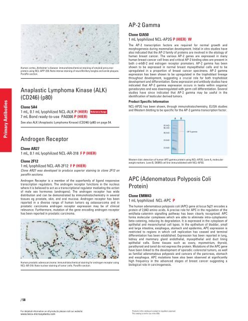
97.4 kD -<br />
66.2 kD -<br />
45.0 kD -<br />
31.0 kD -<br />
21.0 kD -<br />
Western blot: detection of human AP2 gamma protein using NCL-AP2G. Lane A, molecular<br />
weight markers. Lane B, SKBR3 cell line immunoblotted with NCL-AP2G.<br />
APC (Adenomatous Polyposis Coli<br />
Protein)<br />
Clone EMM43<br />
1 mL lyophilized NCL-APC P<br />
The human adenomatous polyposis coli (APC) gene at locus 5q21 encodes a<br />
protein of 2,843 amino acids. A precise role for APC in the regulation of the<br />
wnt/beta-cateninin signalling pathway has been clearly recognized. APC<br />
forms molecular complexes which are able to eliminate intra-cytoplasmic<br />
beta-catening, inducing its degradation. It is expressed in the cytoplasm of<br />
epithelial and mesenchymal cell types. In the epithelium of bladder, small<br />
and large intestine, esophagus, stomach and epidermis, APC expression is<br />
restricted to regions in which cell replication has ceased and terminal<br />
differentiation has been established. Expression has been reported in lung,<br />
kidney and mammary gland endothelial, myoepithelial and duct lining<br />
epithelial cells. Some tissues such as ovary, myometrium, thyroid,<br />
parathyroid and tonsil do not express the protein. Mutations of the APC gene<br />
have been linked to the development of sporadic colorectal tumors, as well<br />
as familial adenomatous polyposis and cancers of the pancreas, stomach<br />
and esophagus. APC mutations have also been observed at significantly<br />
high frequency in the advanced stages of breast cancer suggesting a<br />
biological role in carcinogenesis.<br />
Products in this catalog are subject to regulatory approval.<br />
This catalog is not for use in the USA.<br />
A B<br />
- AP-2 gamma


