

Labelling Review row-Online
Labelling Review row-Online
Labelling Review row-Online

You also want an ePaper? Increase the reach of your titles
YUMPU automatically turns print PDFs into web optimized ePapers that Google loves.
Primary Antibodies<br />
Osteonectin<br />
Clone 15G12<br />
1 mL, 0.1 mL lyophilized NCL-O-NECTIN P (HIER) W<br />
Osteonectin (ON), also known as BM-40 or SPARC (secreted protein, acidic<br />
and rich in cysteine) is a multifunctional glycoprotein (32.5 kD) involved with<br />
tissue mineralization as well as extracellular matrix modelling. ON is the<br />
most abundant glycoprotein secreted by human osteoblasts in developing<br />
bone and odontoblasts of developing teeth. ON mRNA and protein have<br />
been reported to be expressed in non-mineralized tissues such as steroidproducing<br />
cells of the adrenal glands, suprabasal layers of the epidermis,<br />
glomeruli in the kidney, bronchi of the lung, megakaryocytes and large<br />
vessels. This organ-specific distribution of ON in non-mineralized tissues<br />
suggests a role during human development. It is also reported to be<br />
expressed in osteosarcomas, at high levels in osteoblastic osteosarcomas<br />
and at low levels in osteoid formation of the diffuse deposition type.<br />
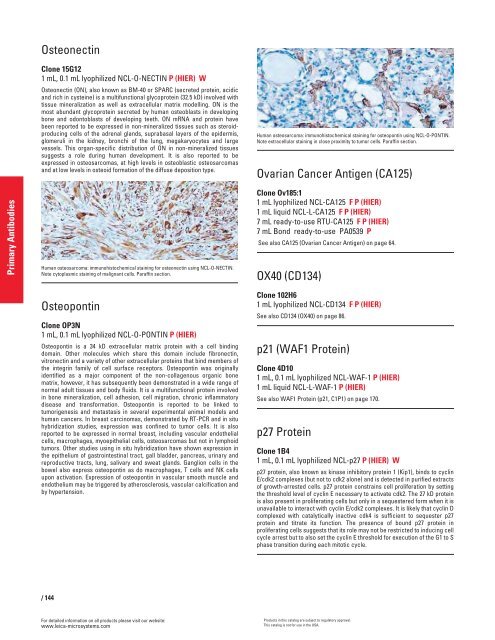
Human osteosarcoma: immunohistochemical staining for osteonectin using NCL-O-NECTIN.<br />
Note cytoplasmic staining of malignant cells. Paraffin section.<br />
Osteopontin<br />
Clone OP3N<br />
1 mL, 0.1 mL lyophilized NCL-O-PONTIN P (HIER)<br />
Osteopontin is a 34 kD extracellular matrix protein with a cell binding<br />
domain. Other molecules which share this domain include fibronectin,<br />
vitronectin and a variety of other extracellular proteins that bind members of<br />
the integrin family of cell surface receptors. Osteopontin was originally<br />
identified as a major component of the non-collagenous organic bone<br />
matrix, however, it has subsequently been demonstrated in a wide range of<br />
normal adult tissues and body fluids. It is a multifunctional protein involved<br />
in bone mineralization, cell adhesion, cell migration, chronic inflammatory<br />
disease and transformation. Osteopontin is reported to be linked to<br />
tumorigenesis and metastasis in several experimental animal models and<br />
human cancers. In breast carcinomas, demonstrated by RT-PCR and in situ<br />
hybridization studies, expression was confined to tumor cells. It is also<br />
reported to be expressed in normal breast, including vascular endothelial<br />
cells, macrophages, myoepithelial cells, osteosarcomas but not in lymphoid<br />
tumors. Other studies using in situ hybridization have shown expression in<br />
the epithelium of gastrointestinal tract, gall bladder, pancreas, urinary and<br />
reproductive tracts, lung, salivary and sweat glands. Ganglion cells in the<br />
bowel also express osteopontin as do macrophages, T cells and NK cells<br />
upon activation. Expression of osteopontin in vascular smooth muscle and<br />
endothelium may be triggered by atherosclerosis, vascular calcification and<br />
by hypertension.<br />
/ 144<br />
For detailed information on all products please visit our website:<br />
www.leica-microsystems.com<br />
Human osteosarcoma: immunohistochemical staining for osteopontin using NCL-O-PONTIN.<br />
Note extracellular staining in close proximity to tumor cells. Paraffin section.<br />
Ovarian Cancer Antigen (CA125)<br />
Clone Ov185:1<br />
1 mL lyophilized NCL-CA125 F P (HIER)<br />
1 mL liquid NCL-L-CA125 F P (HIER)<br />
7 mL ready-to-use RTU-CA125 F P (HIER)<br />
7 mL Bond ready-to-use PA0539 P<br />
See also CA125 (Ovarian Cancer Antigen) on page 64.<br />
OX40 (CD134)<br />
Clone 102H6<br />
1 mL lyophilized NCL-CD134 F P (HIER)<br />
See also CD134 (OX40) on page 86.<br />
p21 (WAF1 Protein)<br />
Clone 4D10<br />
1 mL, 0.1 mL lyophilized NCL-WAF-1 P (HIER)<br />
1 mL liquid NCL-L-WAF-1 P (HIER)<br />
See also WAF1 Protein (p21, C1P1) on page 170.<br />
p27 Protein<br />
Clone 1B4<br />
1 mL, 0.1 mL lyophilized NCL-p27 P (HIER) W<br />
p27 protein, also known as kinase inhibitory protein 1 (Kip1), binds to cyclin<br />
E/cdk2 complexes (but not to cdk2 alone) and is detected in purified extracts<br />
of g<strong>row</strong>th-arrested cells. p27 protein constrains cell proliferation by setting<br />
the threshold level of cyclin E necessary to activate cdk2. The 27 kD protein<br />
is also present in proliferating cells but only in a sequestered form when it is<br />
unavailable to interact with cyclin E/cdk2 complexes. It is likely that cyclin D<br />
complexed with catalytically inactive cdk4 is sufficient to sequester p27<br />
protein and titrate its function. The presence of bound p27 protein in<br />
proliferating cells suggests that its role may not be restricted to inducing cell<br />
cycle arrest but to also set the cyclin E threshold for execution of the G1 to S<br />
phase transition during each mitotic cycle.<br />
Products in this catalog are subject to regulatory approval.<br />
This catalog is not for use in the USA.


