

Labelling Review row-Online
Labelling Review row-Online
Labelling Review row-Online

You also want an ePaper? Increase the reach of your titles
YUMPU automatically turns print PDFs into web optimized ePapers that Google loves.
N-Cadherin<br />
Clone IAR06<br />
1 mL, 0.1 mL liquid NCL-L-N-Cad P (HIER)<br />
N-Cadherin is a member of the cadherin family of calcium dependent cell<br />
adhesion molecules. The classical cadherins include the E, N, R, P and VE-<br />
Cadherins which are believed to be expressed in a tissue specific manner.<br />
The classical cadherins have a characteristic structure comprising an extra<br />
cellular calcium-binding domain, consisting of five repeats, a<br />
transmembrane domain and a highly conserved cytoplasmic domain, which<br />
mediates interactions with cytoskeletal components of the cell via<br />
interactions with intracellular proteins including the catenins. Cadherins<br />
play an important role in cell-cell adhesion, and are implicated in<br />
segregation and aggregation of tissues during development. N-Cadherin is<br />
reported to be expressed in various cell types including neural, myocardial<br />
and mesenchymal cells. During tumor progression increased N-Cadherin<br />
expression concomitant with the loss of E-Cadherin expression is one of the<br />
features of the epithelial to mesenchymal transition, which is associated<br />
with increased tumor invasion and poor prognosis. N-Cadherin has been<br />
proposed as a useful marker in a panel of antibodies to differentiate<br />
mesotheliomas from adenocarcinomas.<br />
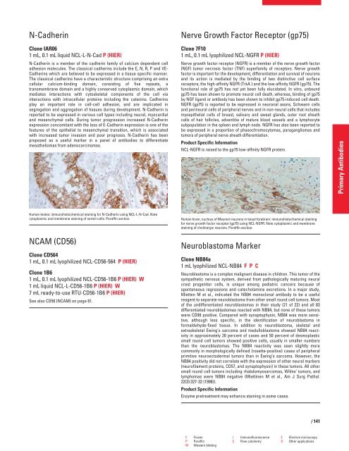
Human testes: immunohistochemical staining for N-Cadherin using NCL-L-N-Cad. Note<br />
cytoplasmic and membrane staining of sertoli cells. Paraffin section.<br />
NCAM (CD56)<br />
Clone CD564<br />
1 mL, 0.1 mL lyophilized NCL-CD56-564 P (HIER)<br />
Clone 1B6<br />
1 mL, 0.1 mL lyophilized NCL-CD56-1B6 P (HIER) W<br />
1 mL liquid NCL-L-CD56-1B6 P (HIER) W<br />
7 mL ready-to-use RTU-CD56-1B6 P (HIER)<br />
See also CD56 (NCAM) on page 81.<br />
Nerve G<strong>row</strong>th Factor Receptor (gp75)<br />
Clone 7F10<br />
1 mL, 0.1 mL lyophilized NCL-NGFR P (HIER)<br />
Nerve g<strong>row</strong>th factor receptor (NGFR) is a member of the nerve g<strong>row</strong>th factor<br />
(NGF) tumor necrosis factor (TNF) superfamily of receptors. Nerve g<strong>row</strong>th<br />
factor is important for the development, differentiation and survival of neurons<br />
and its action is mediated by the binding of two distinctive cell surface<br />
receptors; the high-affinity NGFR (TrkA ) and the low-affinity NGFR (gp75). The<br />
functional role of gp75 has not yet been fully elucidated. In vitro, unbound<br />
gp75 has been shown to promote neural cell death, whereas, binding of gp75<br />
by NGF ligand or antibody has been shown to inhibit gp75-induced cell death.<br />
NGFR (gp75) is reported to be expressed in neuronal axons, Schwann cells<br />
and perineural cells of peripheral nerves and in non-neural cells that includes<br />
myoepithelial cells of breast, salivary and sweat glands, outer root sheath<br />
cells of hair follicles, adventitia of mature blood vessels and a lymphocyte<br />
subpopulation in the spleen and lymph node. NGFR has also been reported to<br />
be expressed in a proportion of phaeochromocytomas, paragangliomas and<br />
tumors of peripheral nerve sheath differentiation.<br />
Product Specific Information<br />
NCL-NGFR is raised to the gp75 low-affinity NGFR protein.<br />
Human brain, nucleus of Meynert neurons in basal forebrain: immunohistochemical staining<br />
for nerve g<strong>row</strong>th factor receptor (gp75) using NCL-NGFR. Note cytoplasmic and membrane<br />
staining of cholinergic neurons. Paraffin section.<br />
Neuroblastoma Marker<br />
Clone NB84a<br />
1 mL lyophilized NCL-NB84 FPC<br />
Neuroblastoma is a complex malignant disease in children. This tumor of the<br />
sympathetic nervous system, derived from pathologically maturing neural<br />
crest progenitor cells, is unique among pediatric cancers because of<br />
spontaneous regressions and catecholamine excretions. In a major study,<br />
Mietten M et al., indicated the NB84 monoclonal antibody to be a useful<br />
reagent to separate neuroblastoma from other small round cell tumors. Most<br />
of the undifferentiated neuroblastomas in their study (21 of 22) and all 83<br />
differentiated neuroblastomas reacted with NB84, but none of these tumors<br />
were CD99 positive. Compared with synaptophysin, NB84 was more sensitive,<br />
although less specific, in the identification of neuroblastoma in<br />
formaldehyde-fixed tissue. In addition to neuroblastoma, skeletal and<br />
extraskeletal Ewing’s sarcoma and medulloblastoma showed NB84 reactivity<br />
in approximately 20 percent of cases and 50 percent of desmoplastic<br />
small round cell tumors showed positive cells, usually in smaller numbers<br />
than the neuroblastomas. The NB84 reactivity was seen slightly more<br />
commonly in morphologically defined (rosette-positive) cases of peripheral<br />
primitive neuroectodermal tumors than in Ewing’s sarcoma. However, the<br />
NB84 positivity did not correlate with the expression of other neural markers<br />
(neurofilament proteins, CD57, and synaptophysin) in these tumors. All other<br />
small round cell tumors including rhabdomyosarcomas, Wilms’ tumors, and<br />
lymphomas were NB84 negative (Miettinen M et al., Am J Surg Pathol.<br />
22(3):327-32 (1998)).<br />
Product Specific Information<br />
Enzyme pretreatment may enhance staining in some cases.<br />
F Frozen I Immunofluorescence E Electron microscopy<br />
P Paraffin C Flow cytometry O Other applications<br />
W Western blotting<br />
/ 141<br />
Primary Antibodies


