

Labelling Review row-Online
Labelling Review row-Online
Labelling Review row-Online

You also want an ePaper? Increase the reach of your titles
YUMPU automatically turns print PDFs into web optimized ePapers that Google loves.
Primary Antibodies<br />
Transforming G<strong>row</strong>th Factor Beta<br />
Clone TGFB17<br />
1 mL, 0.1 mL lyophilized NCL-TGFB P (HIER)<br />
Transforming g<strong>row</strong>th factor beta (TGFB) is a potent cell regulatory<br />
polypeptide homodimer of 25 kD. It variably affects cell g<strong>row</strong>th, differentiation<br />
and other aspects of cellular metabolism such as extracellular matrix<br />
production. Its effect depends upon the cell type and other g<strong>row</strong>th factors<br />
present but in general, TGFB inhibits the g<strong>row</strong>th of epithelial cells and<br />
stimulates the g<strong>row</strong>th of mesenchymal cells. Most breast lesions, benign<br />
and malignant, involve abnormal proliferation and altered architecture of<br />
stromal and/or epithelial elements. Inflammatory cells present in the earliest<br />
lesions of progressive systemic sclerosis (PSS) are reported to release<br />
TGFB possibly resulting in chemotactic recruitment of additional chronic<br />
inflammatory cells. Platelets, a rich source of TGFB, are known to exhibit<br />
aggregability and may contribute to the etiology of PSS.<br />
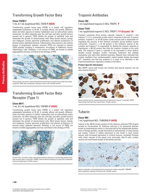
Human breast carcinoma: immunohistochemical staining for transforming g<strong>row</strong>th factor beta<br />
using NCL-TGFB. Note cytoplasmic staining of tumor cells. Paraffin section.<br />
Transforming G<strong>row</strong>th Factor Beta<br />
Receptor (Type 1)<br />
Clone 8A11<br />
1 mL, 0.1 mL lyophilized NCL-TGFBR1 P (HIER)<br />
Transforming g<strong>row</strong>th factor beta (TGFB) is a potent cell regulatory<br />
polypeptide homodimer of 25 kD. It variably affects cell g<strong>row</strong>th, differentiation<br />
and other aspects of cellular metabolism such as extracellular matrix<br />
production. Its effect depends upon the cell type and other g<strong>row</strong>th factors<br />
present but in general, TGFB inhibits the g<strong>row</strong>th of epithelial cells and<br />
stimulates the g<strong>row</strong>th of mesenchymal cells. Most breast lesions, benign<br />
and malignant, involve abnormal proliferation and altered architecture of<br />
stromal and/or epithelial elements. Platelets, a rich source of TGFB, are<br />
known to exhibit aggregability and may contribute to the etiology of PSS.<br />
Human colon, ulcerative colitis: immunohistochemical staining for transforming g<strong>row</strong>th factor<br />
beta receptor (type 1) using NCL-TGFBR1. Note membrane staining of a proportion of epithelial<br />
cells and lymphocytes. Paraffin section.<br />
/ 166<br />
For detailed information on all products please visit our website:<br />
www.leica-microsystems.com<br />
Troponin Antibodies<br />
Clone 1A2<br />
1 mL lyophilized troponin C NCL-TROPC P<br />
Clone T1/61<br />
1 mL lyophilized troponin C NCL-TROPT F P (Enzyme) W<br />
Troponin comprises three protein subunits, troponin C, troponin I and<br />
troponin T. It is a contractile protein which comprises 5 percent of muscle<br />
proteins. Troponin C, an 18 kD protein, binds calcium and is responsible for<br />
regulating the process of thin filament activation during skeletal muscle<br />
contraction. Troponin I, a 21 kD protein, is the inhibitory subunit of the<br />
complex and troponin T is responsible for binding the troponin subunits to<br />
tropomyosin, a 66 kD protein that links the troponin complex to the actin<br />
helix. The troponin C gene is reported to be expressed in three distinct<br />
striated muscle lineages; cardiac myocytes, embryonic fast skeletal<br />
myotubules and adult slow skeletal myocytes. Reports have indicated that<br />
cardiac myofibers from cardiomyopathic rodent models display decreased<br />
Ca2+ sensitivity and that this property is a result of an alteration in the<br />
troponin/tropomyosin regulatory complex in the fibers.<br />
Product Specific Information<br />
NCL-TROPT reacts with human and chicken fast muscle troponin, but not<br />
slow muscle troponin T.<br />
Human skeletal muscle: immunohistochemical staining for troponin T using NCL-TROPT.<br />
Note intense staining of fast muscle fibers. Paraffin section.<br />
Tuberin<br />
Clone 3B4<br />
1 mL lyophilized NCL-TUBERIN P (HIER)<br />
Tuberin is the 180 kD protein product of the tuberous sclerosis (TSC-2) gene<br />
located on chromosome 16. Tuberous sclerosis is an autosomal disorder<br />
known as phakomatosis which is characterized by the widespread development<br />
of benign g<strong>row</strong>ths, usually described as hamartomata, in many tissues<br />
and organs. Tuberin is widely expressed at low levels in most human<br />
tissues, with increased expression in cortical neurons, cerebellar Purkinje<br />
cells, motor neurons of the spinal cord, pancreatic islet B cells, cardiac<br />
muscle and small blood vessels of the kidney and skin.<br />
Human cerebellum: immunohistochemical staining for tuberin using NCL-TUBERIN.<br />
Note cytoplasmic staining of Purkinje cells. Paraffin section.<br />
Products in this catalog are subject to regulatory approval.<br />
This catalog is not for use in the USA.


