

Labelling Review row-Online
Labelling Review row-Online
Labelling Review row-Online

You also want an ePaper? Increase the reach of your titles
YUMPU automatically turns print PDFs into web optimized ePapers that Google loves.
Primary Antibodies<br />
MAGE-1<br />
Clone 6C1<br />
1 mL lyophilized NCL-MAGE-1 P (HIER)<br />
The human MAGE gene products are recognised by major<br />
histocompatability complex-restricted cytotoxic T lymphocytes. MAGE-1,<br />
also known as tumor rejection antigen, is a target for immunotherapy in<br />
patients with hepatocellular carcinoma (HCC). MAGE-1 is reported to be<br />
expressed in about 60 per cent of HCC cases. Other studies utilising reverse<br />
transcriptase-PCR and southern blot hybridisation techniques have reported<br />
MAGE genes to be expressed in malignant tumors and pre-cancerous<br />
lesions but not in benign tumors.<br />
Maspin<br />
Clone EAW24<br />
1 mL, 0.1 mL lyophilized NCL-MASPIN P (HIER)<br />
Maspin, or mammary-specific serpin, is a tumor suppressor protein of 42 kD<br />
that belongs to the serine proteinase inhibitor (serpin) family. It is reported to be<br />
expressed in normal breast and prostatic epithelial cells but is downregulated<br />
in carcinomas derived from these cell types. The expression of maspin is<br />
controlled at the transcriptional level by a combination of elements including<br />
Ets, AP-1 and p53. The tumor suppressor activity of maspin may depend on its<br />
ability to inhibit angiogenesis. In breast myoepithelial cells, maspin is<br />
predominantly a soluble cytoplasmic protein which associates with secretory<br />
vesicles and is present at the cell surface. The loss of maspin in breast tumors<br />
is reported to be a progressive process and expression decreases with<br />
increasing malignancy of primary tumors and is absent from lymph node and<br />
distant metastases. In rats, maspin mRNA has been detected in mammary<br />
gland, vagina, bladder, thymus, small intestine, ventral prostate, seminal<br />
vesicles and thyroid, but is absent from heart, lung, liver, brain and kidney.<br />
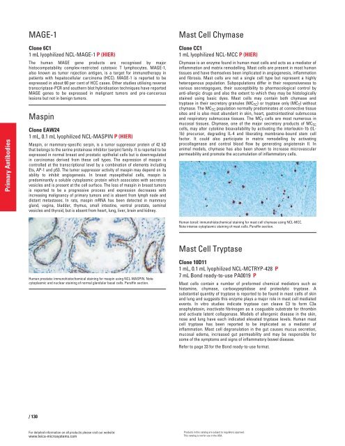
Human prostate: immunohistochemical staining for maspin using NCL-MASPIN. Note<br />
cytoplasmic and nuclear staining of normal glandular basal cells. Paraffin section.<br />
/ 130<br />
For detailed information on all products please visit our website:<br />
www.leica-microsystems.com<br />
Mast Cell Chymase<br />
Clone CC1<br />
1 mL lyophilized NCL-MCC P (HIER)<br />
Chymase is an enzyme found in human mast cells and acts as a mediator of<br />
inflammation and matrix remodelling. Mast cells are present in most human<br />
tissues and have themselves been implicated in angiogenesis, inflammation<br />
and fibrosis. Mast cells are not a single cell type but represent a highly<br />
heterogenous population. Subpopulations differ in their responsiveness to<br />
various secretagogues, their susceptibility to pharmacological control by<br />
anti-allergic drugs and also the extent to which they may be histologically<br />
stained using basic dyes. Mast cells may contain both chymase and<br />
tryptase in their secretory granules (MC TC ) or tryptase only (MC T ) without<br />
chymase. The MC TC population normally predominates at connective tissue<br />
sites and is also most abundant in skin, heart, gastrointestinal submucosa<br />
and respiratory submucosa tissues. The MC T cells are most numerous in<br />
mucosal tissues. Chymase, one of the major secretory products of MC TC<br />
cells, may alter cytokine bioavailability by activating the interleukin-1b (IL-<br />
1b) precursor, degrading IL-4 and liberating membrane-bound stem cell<br />
factor. It could also participate in matrix remodelling by activating<br />
procollagenase and control blood flow by generating angiotensin II. In<br />
animal models, chymase has also been shown to increase microvascular<br />
permeability and promote the accumulation of inflammatory cells.<br />
Human tonsil: immunohistochemical staining for mast cell chymase using NCL-MCC.<br />
Note intense cytoplasmic staining of mast cells. Paraffin section.<br />
Mast Cell Tryptase<br />
Clone 10D11<br />
1 mL, 0.1 mL lyophilized NCL-MCTRYP-428 P<br />
7 mL Bond ready-to-use PA0019 P<br />
Mast cells contain a number of preformed chemical mediators such as<br />
histamine, chymase, carboxypeptidase and proteolytic tryptase. A<br />
substantial quantity of tryptase is reported to be found in mast cells of skin<br />
and lung and suggests this enzyme plays a major role in mast cell mediated<br />
events. In vitro studies indicate tryptase can cleave C3 to form C3a<br />
anaphylatoxin, inactivate fibrinogen as a coaguable substrate for thrombin<br />
and activate latent collagenase. Models of allergenic disease in the skin,<br />
nose and lung have each indicated elevated tryptase levels. Human mast<br />
cell tryptase has been reported to be implicated as a mediator of<br />
inflammation. Mast cell degranulation in the gut causes mucus secretion,<br />
mucosal edema, increased gut permeability and may be responsible for<br />
some of the symptoms and signs of inflammatory bowel disease.<br />
Refer to page 33 for the Bond ready-to-use format.<br />
Products in this catalog are subject to regulatory approval.<br />
This catalog is not for use in the USA.


