

Labelling Review row-Online
Labelling Review row-Online
Labelling Review row-Online

Create successful ePaper yourself
Turn your PDF publications into a flip-book with our unique Google optimized e-Paper software.
Primary Antibodies<br />
CA19-9 (Sialyl Lewis a )<br />
Clone C241:5:1:4<br />
1 mL lyophilized NCL-CA19-9 F P (HIER)<br />
1 mL liquid NCL-L-CA19-9 F P (HIER)<br />
7 mL Bond ready-to-use PA0424 P (HIER)<br />
CA19-9 is an epitope on the sialylated Lewis a carbohydrate structure.<br />
Sialylated Lewis a plays a role in cell adhesion by acting as a functional<br />
ligand for the inducible adhesion molecule E-selectin. CA19-9 and CA50<br />
(carcinoma associated mucin antigen) are useful serum markers in the<br />
diagnosis and follow up of gastrointestinal and pancreatic cancers. In<br />
carcinoma of the pancreas, it is reported that the immunohistochemical<br />
expression of both CA19-9 and CA50 correlates with tumor differentiation<br />
where the strongest staining is observed in well differentiated tumors.<br />
These two markers are also reported in a number of benign lesions such as<br />
chronic pancreatitis.<br />
Product Specific Information<br />
Clone C241:5:1:4 reacts specifically with Sialyl Lewisa - containing glycolipids,<br />
showing no crossreaction with Lewisa , Lewisb , or other structurally related<br />
molecules. The epitope recognized by NCL-L-CA19-9 is designated CA19-9<br />
and is similar to CA50 (carcinoma associated mucin antigen).<br />
Refer to page 16 for the Bond ready-to-use format.<br />
Normal human colon: immunohistochemical staining for Sialyl Lewis a antigen using<br />
NCL-L-CA19-9. Note extracellular-associated staining of colonic epithelial cells. Paraffin<br />
section.<br />
CA125 (Ovarian Cancer Antigen)<br />
Clone Ov185:1<br />
1 mL lyophilized NCL-CA125 F P (HIER)<br />
1 mL liquid NCL-L-CA125 F P (HIER)<br />
7 mL ready-to-use RTU-CA125 F P (HIER)<br />
7 mL Bond ready-to-use PA0539 P (HIER)<br />
CA125 antigen is usually associated with ovarian epithelial malignancies.<br />
Serum assays are widely used to detect this protein in the monitoring of<br />
ovarian cancers. CA125 antigen may also be detected by immunohistochemistry<br />
and expression has been found in neoplasms such as<br />
seminal vesicle carcinoma and anaplastic lymphoma. CA125 antigen is not<br />
found exclusively in malignant tumors. CA125 is also known as MUC16.<br />
Refer to page 16 for the Bond ready-to-use format.<br />
/64<br />
For detailed information on all products please visit our website:<br />
www.leica-microsystems.com<br />
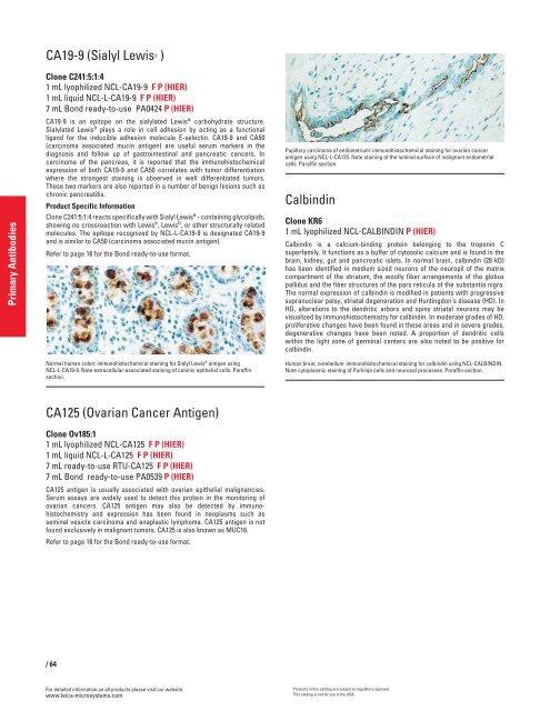
Papillary carcinoma of endometrium: immunohistochemical staining for ovarian cancer<br />
antigen using NCL-L-CA125. Note staining of the luminal surface of malignant endometrial<br />
cells. Paraffin section.<br />
Calbindin<br />
Clone KR6<br />
1 mL lyophilized NCL-CALBINDIN P (HIER)<br />
Calbindin is a calcium-binding protein belonging to the troponin C<br />
superfamily. It functions as a buffer of cytosolic calcium and is found in the<br />
brain, kidney, gut and pancreatic islets. In normal brain, calbindin (28 kD)<br />
has been identified in medium sized neurons of the neuropil of the matrix<br />
compartment of the striatum, the woolly fiber arrangements of the globus<br />
pallidus and the fiber structures of the pars reticula of the substantia nigra.<br />
The normal expression of calbindin is modified in patients with progressive<br />
supranuclear palsy, striatal degeneration and Huntingdon's disease (HD). In<br />
HD, alterations to the dendritic arbors and spiny striatal neurons may be<br />
visualized by immunohistochemistry for calbindin. In moderate grades of HD,<br />
proliferative changes have been found in these areas and in severe grades,<br />
degenerative changes have been noted. A proportion of dendritic cells<br />
within the light zone of germinal centers are also noted to be positive for<br />
calbindin.<br />
Human brain, cerebellum: immunohistochemical staining for calbindin using NCL-CALBINDIN.<br />
Note cytoplasmic staining of Purkinje cells and neuronal processes. Paraffin section.<br />
Products in this catalog are subject to regulatory approval.<br />
This catalog is not for use in the USA.


