

Labelling Review row-Online
Labelling Review row-Online
Labelling Review row-Online

Create successful ePaper yourself
Turn your PDF publications into a flip-book with our unique Google optimized e-Paper software.
Calcitonin<br />
Clone CL1948 New!<br />
1 mL, 0.1 mL liquid NCL-L-CALCITONIN P (Enzyme)<br />
Polyclonal<br />
0.5 mL lyophilized NCL-CALp FP<br />
7 mL Bond ready-to-use PA0406 P (Enzyme)<br />
Calcitonin (CT) is a 32 amino acid peptide synthesized by the parafollicular C<br />
cells of the thyroid. It acts through its receptors to inhibit osteoclast<br />
mediated bone resorption, decrease calcium resorption by the kidney and<br />
decrease calcium absorption by the intestines. The action of calcitonin is<br />
therefore to cause a reduction in serum calcium, an effect opposite to that<br />
of parathyroid hormone. The calcitonin gene transcript also encodes the<br />
calcitonin gene-related peptide (CGRP), which is thought to be a potent<br />
vasodilator. The tissue specificity of the transcript produced depends on<br />
alternative splicing of the CT/CGRP gene transcript. In the parafollicular<br />
cells of the thyroid 95 percent of the CT/CGRP is processed and translated to<br />
produce CT, however, in neuronal cells 99 percent of the CT/CGRP RNA is<br />
translated into CGRP. The C cells of the thyroid give rise to an endocrine<br />
tumor, medullary thyroid carcinoma (MTC), which occurs in a sporadic (75<br />
percent of cases) and hereditary form (25 percent of cases). Familial MTC is<br />
associated with C cell hyperplasia (CCH), whereas sporadic MTC is thought<br />
not to be. However, in the general population CCH is present in 20-30<br />
percent of thyroid glands, either with normal histology, thyroiditis or<br />
follicular tumors.<br />
Refer to page 16 for the Bond ready-to-use format.<br />
Human medullary thyroid carcinoma: immunohistochemical staining for calcitonin using<br />
NCL-L- CALCITONIN. Paraffin section.<br />
Calmodulin<br />
Clone 6312<br />
1 mL lyophilized NCL-CALMODULIN P (HIER)<br />
Calmodulin, a ubiquitous eukaryotic calcium binding protein, is a principal<br />
mediator of the calcium signal. It participates in signalling pathways<br />
inducing proliferation, motility and cell cycle progression. Human calmodulin<br />
is encoded by three genes CALM1, CALM2 and CALM3 located on different<br />
chromosomes. The vertebrate CALM family of genes is unique in that its<br />
members specify an identical protein. The protein itself is made up of 148<br />
amino acids and has four calcium binding domains. As calmodulin is<br />
essential for normal cell function, it is likely that levels are tightly controlled<br />
both temporally and spatially. Immunohistochemical staining for calmodulin<br />
has been reported in the epithelia of testis, breast, stomach, prostate, gall<br />
bladder as well as in macrophages, fibroblasts and sebaceous glands within<br />
the dermis of skin. In healing skin wounds, calmodulin is found at its highest<br />
levels in maturing keratinocytes. It is noticeably abundant in epidermis close<br />
to a wound and re-epithelializing margins where calcium levels are highest.<br />
In studies of Alzheimer's brains, calmodulin immunostaining has been<br />
reported to be lost in cortical regions where large amounts of aluminium<br />
have accumulated.<br />
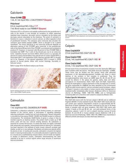
Human in situ ductal carcinoma of breast: immunohistochemical staining for calmodulin using<br />
NCL-CALMODULIN. Note cytoplasmic and membrane staining of malignant cells. Paraffin<br />
section.<br />
Calpain<br />
Clone Calp3d/2C4<br />
2.5 mL lyophilized NCL-CALP-2C4 W<br />
Clone Calp3c/11B3<br />
2.5 mL, 1mL lyophilized NCL-CALP-11B3 W<br />
Clone Calp3c/12A2<br />
2.5 mL, 1 mL lyophilized NCL-CALP-12A2 W<br />
At least seven forms of autosomal recessive muscular dystrophy (MD) have<br />
been included under the banner “limb girdle muscular dystrophy” (LGMD).<br />
These forms may be divided into two groups; those with abnormal<br />
expression of the dystrophin/glycoprotein complex and those in which<br />
labeling of the proteins in this complex is unaffected. Thus the<br />
sarcoglycanopathies (also known as LGMD types 2C, 2D, 2E and 2F) are<br />
caused by defects in the genes for gamma, alpha, beta and deltasarcoglycan<br />
on chromosomes 13q12, 17q21, 4q12 and 5q33, respectively.<br />
Among the dystrophies in which expression of the sarcoglycans is normal,<br />
the gene responsible for LGMD2A has been identified as the chromosome<br />
15q15-encoded muscle-specific calcium-activated neutral protease, calpain<br />
3. Calpain 3 enzyme is only stable in human muscle when homogenized in<br />
treatment buffer immediately after harvest. (Anderson LVB et al. American<br />
Journal of Pathology. 153(4): 1169-1179 (1998)), and in homogenates<br />
containing SDS and is therefore well suited for analysis by Western blot.<br />
Product Specific Information<br />
NCL-CALP-2C4 reacts with the full-size calpain 3 (94 kD) and an additional<br />
fragment (30 kD) in human skeletal muscle. NCL-CALP-12A2 reacts with fullsize<br />
protein plus apparent degradation products at approximately 60 kD.<br />
Specificity of these antibodies has been confirmed by the loss of all these<br />
bands in samples with null gene mutations. NCL-CALP-11B3 reacts with<br />
calpain 3 bands at 94 and 60 kD, pre- and post-autolyzed forms of the<br />
ubiquitous calpains 1 and 2 (� and m-calpain) staining a group of bands<br />
between 76 and 84 kD in human skeletal muscle (Anderson LVB et al.<br />
American Journal of Pathology. 153(4): 1169-1179, (1998)) Cross-reactivities in<br />
different animals and tissues are described (see reference).<br />
F Frozen I Immunofluorescence E Electron microscopy<br />
P Paraffin C Flow cytometry O Other applications<br />
W Western blotting<br />
/65<br />
Primary Antibodies


