

Labelling Review row-Online
Labelling Review row-Online
Labelling Review row-Online

You also want an ePaper? Increase the reach of your titles
YUMPU automatically turns print PDFs into web optimized ePapers that Google loves.
Human appendix: immunohistochemical staining for mast cell tryptase using NCL-MCTRYP-<br />
428. Note cytoplasmic staining of mast cells. Paraffin section.<br />
Matrix Metalloproteinase Antibodies<br />
Clone 17B11<br />
1 mL, 0.1 mL lyophilized Matrix Metalloproteinase 2<br />
NCL-MMP2-507 P (HIER)<br />
Clone 15W2<br />
1 mL, 0.1 mL lyophilized Matrix Metalloproteinase 9<br />
NCL-MMP9-439 FP<br />
Clone 5E4<br />
1 mL lyophilized Matrix Metalloproteinase 10<br />
NCL-MMP10 P (HIER)<br />
Clone 9F6<br />
1 mL lyophilized Matrix Metalloproteinase 19<br />
NCL-MMP19 P<br />
The matrix metalloproteinases (MMPs) are a family of zinc-containing<br />
enzymes, which are involved in the degradation of different components of<br />
the extracellular matrix and tissue remodelling. MMPs are expressed widely<br />
during g<strong>row</strong>th and development. The MMPs have been classified into<br />
collagenases, gelatinases and stromelysins, based on the in vitro substrate<br />
specificity. More recently, several MMPs have been identified as<br />
membrane-type specific and matrilysin families. MMPs are multidomain<br />
proteins and are secreted as inactive precursors which are activated by<br />
cleavage of an N-terminal pro-peptide. The major natural inhibitors of<br />
MMPs are tissue inhibitors of matrix metalloproteinases (TIMPs) which<br />
complex with MMPs and are involved in regulating the activity and<br />
activation of individual MMPs. MMP2 (also known as gelatinase A) is able to<br />
initiate degradation of type IV collagen. MMP9 degrades collagen type IV, a<br />
major component of extracellular matrix. MMP9 is also reported to be<br />
expressed in normal kidney tubules, hepatocytes, spermatids, myocytes,<br />
stomach parietal cells, prostatic columnar epithelium and uterine cells.<br />
MMP10 is also known as stromelysin-2 and has a wide range of substrates<br />
including proteoglycan, laminin, fibronectin, collagen IV, collagen IX and the<br />
telopeptides of other collagens. However, some of the more recently<br />
identified MMPs, such as MMP19 - which cleaves aggrecan and cartilage<br />
oligomeric protein, and has several novel structural features, do not fall into<br />
these traditional groupings. MMP19 is reported to be expressed mainly in<br />
placenta, lung, pancreas, ovary, spleen, intestine, breast tissue, smooth<br />
muscle, capillary walls and the endothelial layers of large and medium sized<br />
blood vessels.<br />
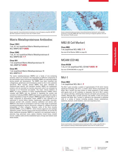
Human endometrial adenocarcinoma: immunohistochemical staining for matrix metalloproteinase<br />
2 using NCL-MMP2-507. Note intense staining of malignant epithelial cells and<br />
their secretions. Paraffin section.<br />
MB2 (B Cell Marker)<br />
Clone MB2<br />
1 mL lyophilized NCL-MB2 FP<br />
See also B Cell Marker (MB2) on page 59.<br />
MCAM (CD146)<br />
Clone N1238<br />
1 mL, 0.1 mL lyophilized NCL-CD146 P (HIER) W<br />
See also CD146 (MCAM) on page 87.<br />
Mcl-1<br />
Clone 38G3<br />
1 mL lyophilized NCL-Mcl-1 P<br />
The Mcl-1 gene encodes a protein of approximately 37 kD which shares<br />
significant homology with bcl-2, a protein which blocks programmed cell<br />
death. Mcl-1 protein has been shown to inhibit apoptosis in cells treated<br />
with agents such as UV irradiation or etoposide. Like bcl-2, Mcl-1 protein<br />
can interact with Bax, inhibiting Bax-induced cell death and promoting cell<br />
viability. Although Mcl-1 protein prolongs cell viability, it does not prevent<br />
eventual cell death. Mcl-1 protein is reported to be expressed in epithelial<br />
cells in a variety of tissues including prostate, breast, endometrium,<br />
epidermis, stomach, small intestine, colon and respiratory tract.<br />
Human small intestine: immunohistochemical staining for Mcl-1 protein using NCL-Mcl-1.<br />
Note cytoplasmic staining of differentiated epithelial cells of the villi. Paraffin section.<br />
F Frozen I Immunofluorescence E Electron microscopy<br />
P Paraffin C Flow cytometry O Other applications<br />
W Western blotting<br />
/ 131<br />
Primary Antibodies


