

Labelling Review row-Online
Labelling Review row-Online
Labelling Review row-Online

You also want an ePaper? Increase the reach of your titles
YUMPU automatically turns print PDFs into web optimized ePapers that Google loves.
Calretinin<br />
Clone CAL6<br />
7 mL Bond ready-to-use PA0346 P (HIER)<br />
For In Vitro Diagnostic Use<br />
Calretinin is a calcium-binding protein of 29 kD, reported to be abundantly<br />
expressed in neurons. Outside the nervous system, calretinin expression<br />
has been demonstrated in a range of cell types including mesothelial cells,<br />
steroid producing cells, some neuroendocrine cells, eccrine sweat glands<br />
and other cell types. The presence of calretinin is reported to be a useful<br />
marker primarily for two purposes: differentiating malignant mesothelioma<br />
from carcinomas; and the differential characterization of ovarian stromal<br />
tumors. Calretinin-positive cells have also been reported in the convoluted<br />
tubules of kidney with some antibodies.<br />
Product Specific Information<br />
Bond ready-to-use Calretinin (CAL6) is recommended for use as part of an<br />
antibody panel for the identification of mesothelioma.<br />
Also available as a Novocastra concentrate, refer to page 66.<br />
Mesothelioma: immunohistochemical staining with Bond ready-to-use Calretinin (CAL6) using<br />
Bond Polymer Refine Detection.<br />
Carcinoembryonic Antigen<br />
Clone II-7<br />
7 mL Bond ready-to-use PA0004 P (HIER)<br />
For In Vitro Diagnostic Use<br />
Carcinoembryonic antigen (CEA) is a heterogeneous cell surface<br />
glycoprotein produced by cells of fetal colon. Low levels are also found on<br />
normal mucosal epithelia of the adult colon and a variety of other normal<br />
tissues. CEA is encoded by the CEA gene that is located on chromosome 19.<br />
It is a member of the CEA gene family, which in turn is a subfamily of the<br />
immunoglobulin superfamily. Cell adhesion properties are now well<br />
recognized for CEA. It is believed that the expression of this glycoprotein in<br />
conjunction with other known adhesion molecules will influence the cellcell<br />
interaction.<br />
Also available as a Novocastra concentrate, refer to page 67.<br />
Colon adenocarcinoma: immunohistochemical staining with Bond ready-to-use<br />
Carcinoembryonic Antigen (II-7) using Bond Polymer Refine Detection.<br />
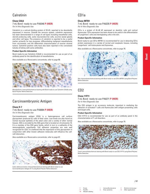
CD1a<br />
Clone MTB1<br />
7 mL Bond ready-to-use PA0235 P (HIER)<br />
For In Vitro Diagnostic Use<br />
CD1a is a protein of 43–49 kD expressed on dendritic cells and cortical<br />
thymocytes. CD1a expression has been shown to be useful in the differentiation<br />
of Langerhans’ cells and interdigitating cells in the skin.<br />
Product Specific Information<br />
Bond ready-to-use CD1a (MTB1) is recommended for use in detecting CD1a<br />
protein expression in a variety of normal and neoplastic tissues, including<br />
Langerhans' cell histiocytosis and thymomas.<br />
Also available as a Novocastra concentrate, refer to page 70.<br />
Skin: immunohistochemical staining with Bond ready-to-use CD1a (MTB1) using Bond Polymer<br />
Refine Detection.<br />
CD2<br />
Clone 11F11<br />
7 mL Bond ready-to-use PA0271 P (HIER)<br />
For In Vitro Diagnostic Use<br />
The CD2 antigen is an accessory molecule, important in mediating the<br />
adhesion of activated T cells and thymocytes with antigen-presenting cells<br />
and target cells.<br />
Product Specific Information<br />
CD2 (11F11) is recommended for use as part of an antibody panel in the<br />
characterization of T cell disorders.<br />
Also available as a Novocastra concentrate, refer to page 70.<br />
Tonsil: immunohistochemical staining with Bond ready-to-use CD2 (11F11) using Bond Polymer<br />
Refine Detection.<br />
F Frozen I Immunofluorescence E Electron microscopy<br />
P Paraffin C Flow cytometry O Other applications<br />
W Western blotting<br />
/17<br />
Bond


