

Labelling Review row-Online
Labelling Review row-Online
Labelling Review row-Online

Create successful ePaper yourself
Turn your PDF publications into a flip-book with our unique Google optimized e-Paper software.
Alpha-1-Antichymotrypsin<br />
Polyclonal<br />
1 mL lyophilized NCL-A1ACp P<br />
Alpha-1-Antichymotrypsin (ACT) is an early-stage acute phase protein and a<br />
member of the serine proteinase inhibitor or serpin superfamily. The precise role<br />
of ACT is uncertain but it is thought to act as an anti-inflammatory agent<br />
inhibiting chymotrypsin, cathepsin G, mast cell chymase, neutrophil chemotaxis<br />
and superoxide anion production. ACT is synthesized primarily by hepatocytes of<br />
the liver. Lower levels of synthesis have also been discovered via<br />
immunohistochemical analysis in mast cells, endothelial cells, breast and<br />
intestinal epithelial cells. ACT also exists in the brain. Research has shown that it<br />
is found in amyloid fibrils, endothelial cells and the cytoplasm of astroglial cells<br />
in certain brain abnormalities. Further research has also shown that a major<br />
proportion of prostate-specific antigen (PSA) in serum exists complexed to ACT.<br />
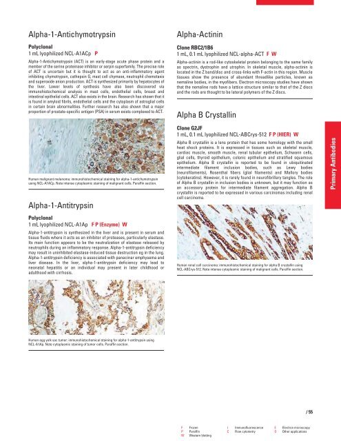
Human malignant melanoma: immunohistochemical staining for alpha-1-antichymotrypsin<br />
using NCL-A1ACp. Note intense cytoplasmic staining of malignant cells. Paraffin section.<br />
Alpha-1-Antitrypsin<br />
Polyclonal<br />
1 mL lyophilized NCL-A1Ap F P (Enzyme) W<br />
Alpha-1-antitrypsin is synthesized in the liver and is present in serum and<br />
tissue fluids where it acts as an inhibitor of proteases, particularly elastase.<br />
Its main function appears to be the neutralization of elastase released by<br />
neutrophils during an inflammatory response. Alpha-1-antitrypsin deficiency<br />
may result in uninhibited elastase-induced tissue destruction eg in the lung.<br />
Alpha-1-antitrypsin deficiency is associated with panacinar emphysema and<br />
liver disease. In the liver, alpha-1-antitrypsin deficiency may lead to<br />
neonatal hepatitis or an individual may present in later childhood or<br />
adulthood with cirrhosis.<br />
Human egg yolk sac tumor: immunohistochemical staining for alpha-1-antitrypsin using<br />
NCL-A1Ap. Note cytoplasmic staining of tumor cells. Paraffin section.<br />
Alpha-Actinin<br />
Clone RBC2/1B6<br />
1 mL, 0.1 mL lyophilized NCL-alpha-ACT FW<br />
Alpha-actinin is a rod-like cytoskeletal protein belonging to the same family<br />
as spectrin, dystrophin and utrophin. In skeletal muscle, alpha-actinin is<br />
located in the Z band/disc and cross-links with F-actin in this region. Muscle<br />
tissues show the presence of abundant threadlike particles, known as<br />
nemaline bodies, in the myofibers. Electron microscopy studies have shown<br />
that the nemaline rods have a lattice structure similar to that of the Z discs<br />
and the rods are thought to be lateral polymers of the Z discs.<br />
Alpha B Crystallin<br />
Clone G2JF<br />
1 mL, 0.1 mL lyophilized NCL-ABCrys-512 F P (HIER) W<br />
Alpha B crystallin is a lens protein that has some homology with the small<br />
heat shock proteins. It is expressed in tissues such as skeletal muscle,<br />
cardiac muscle, smooth muscle, renal tubular epithelium, Schwann cells,<br />
glial cells, thyroid epithelium, colonic epithelium and stratified squamous<br />
epithelium. Alpha B crystallin is reported to be found in ubiquitinated<br />
intermediate filament inclusion bodies, such as Lewy bodies<br />
(neurofilaments), Rosenthal fibers (glial filaments) and Mallory bodies<br />
(cytokeratins). However, it is rarely found in neurofibrillary tangles. The role<br />
of Alpha B crystallin in inclusion bodies is unknown, but it may function as<br />
an accessory protein for intermediate filament aggregation. Alpha B<br />
crystallin is reported to be expressed in various carcinomas including renal<br />
cell carcinoma.<br />
Human renal cell carcinoma: immunohistochemical staining for alpha B crystallin using<br />
NCL-ABCrys-512. Note intense cytoplasmic staining of malignant cells. Paraffin section.<br />
F Frozen I Immunofluorescence E Electron microscopy<br />
P Paraffin C Flow cytometry O Other applications<br />
W Western blotting<br />
/55<br />
Primary Antibodies


