

Labelling Review row-Online
Labelling Review row-Online
Labelling Review row-Online

You also want an ePaper? Increase the reach of your titles
YUMPU automatically turns print PDFs into web optimized ePapers that Google loves.
Complement Component C9<br />
Clone 10A6<br />
1 mL, 0.1 mL lyophilized NCL-CCC9 P (HIER)<br />
Complement component C9 binds to the C5b-8 complex as the final protein<br />
of the membrane attack complex. After binding, it undergoes a conformational<br />
change and inserts itself into the cell membrane, forming<br />
transmembrane channels. Complement component C9 acts in a similar way<br />
to perforin, a pore forming protein found in cytotoxic T cells. Male and<br />
female reproductive tissues express and synthesize complement<br />
components, binding proteins and receptors, although the implications of<br />
this is unclear. The detection of complement component C9 has been<br />
reported in cases of acute myocardial damage at necropsy. Detection of<br />
myocardial infarction or diffuse damage can be unreliable with conventional<br />
methods of examination of the heart such as enzyme histochemistry or by<br />
the elaborate technique of quantification of contraction band necrosis.<br />
Human myocardium: immunohistochemical staining for complement component C9 using<br />
NCL-CCC9. Note staining of partially necrotic myocardium and vessel walls. Paraffin section.<br />
CPP32 (Caspase-3)<br />
Clone JHM62<br />
1 mL, 0.1 mL lyophilized NCL-CPP32 P (HIER) W<br />
Cysteine protease protein (CPP)-32 is a member of the interleukin-1 betaconverting<br />
enzyme (ICE) family of mammalian proteases which specifically<br />
cleaves substrates at the C-terminal side of aspartic acid residues.<br />
Members of this family have been implicated in apoptosis and CPP32<br />
(caspase-3) is thought to act as a control mediator of programmed cell<br />
death in mammalian cells. CPP32 is synthesized as an inactive 32 kD<br />
proenzyme and is processed during apoptosis to its active form which is<br />
responsible for the cleavage of poly (ADP-ribose) polymerase (PARP), actin<br />
and sterol regulatory element binding protein (SREBP). CPP32 is reported to<br />
be found in epithelial cells of skin, renal proximal tubules and collecting<br />
ducts, epithelioreticular cells of the thymus and bronchial, colonic and<br />
salivary duct epithelia. Chondrocytes, bone osteocytes, megakaryocytes,<br />
mature neutrophils of bone mar<strong>row</strong> and plasma cells of the tonsil, lymph<br />
node and bone mar<strong>row</strong> are also reported to express CPP32 antigen.<br />
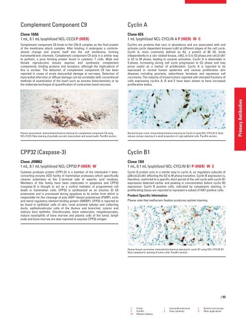
Cyclin A<br />
Clone 6E6<br />
1 mL lyophilized NCL-CYCLIN A P (HIER) W C<br />
Cyclins are proteins that vary in abundance and are associated with and<br />
activate cyclin dependent kinases (cdk) at different stages of the cell cycle.<br />
Cyclin A, more commonly defined as A2, a protein of 60 kD, binds<br />
independently to a cdc-related kinase, cdk2, in S to G2 phase and cdc2/cdk1<br />
in G2 to M phase, leading to enzyme activation. Cyclin A is detectable in<br />
S phase, increasing during cell cycle progression to G2 phase and may<br />
prove useful as a marker of proliferation. Cyclin A is reported to be<br />
expressed in normal human epidermis and various proliferative skin<br />
diseases including psoriasis, seborrhoeic keratosis and squamous cell<br />
carcinoma. The majority of breast tumors reported with elevated fractions of<br />
cells expressing cyclins A, B and E have been shown to have increased<br />
proliferative status.<br />
Normal human colon: immunohistochemical staining for Cyclin A using NCL-CYCLIN A. Note<br />
intense nuclear staining of a small proportion of crypt epithelial cells. Paraffin section.<br />
Cyclin B1<br />
Clone 7A9<br />
1 mL, 0.1 mL lyophilized NCL-CYCLIN B1 P (HIER) W C<br />
Cyclin B protein acts in a similar way to cyclin A, as regulatory subunits of<br />
p34/cdc2/cdk1 affecting the G2 to M phase transition. Cyclin B expression is,<br />
therefore, restricted to a specific short period of the cell cycle with cyclin B1<br />
expression detected earlier and peaking in concentration before cyclin B2<br />
expression. Cyclin B positive cells, indicated by cytoplasmic staining, in<br />
proliferating tissue are reported to represent a subset of Ki67 positive cells.<br />
Product Specific Information<br />
Please note that methacarn fixation produces optimal staining.<br />
Human breast carcinoma: immunohistochemical staining for cyclin B1 using NCL-CYCLIN B1.<br />
Note cytoplasmic staining of tumor cells. Paraffin section.<br />
F Frozen I Immunofluorescence E Electron microscopy<br />
P Paraffin C Flow cytometry O Other applications<br />
W Western blotting<br />
/93<br />
Primary Antibodies


