

Labelling Review row-Online
Labelling Review row-Online
Labelling Review row-Online

You also want an ePaper? Increase the reach of your titles
YUMPU automatically turns print PDFs into web optimized ePapers that Google loves.
Primary Antibodies<br />
c-erbB-3 Oncoprotein<br />
Clone RTJ1<br />
1 mL lyophilized NCL-c-erbB-3 F P (HIER) O<br />
The c-erbB-3 oncoprotein is a member of the type 1 g<strong>row</strong>th factor receptor<br />
family which also includes c-erbB-2 and epidermal g<strong>row</strong>th factor receptor<br />
(EGFR). These receptors share a common overall structure consisting of an<br />
extracellular domain, a transmembrane region and a cytoplasmic domain.<br />
The expression of c-erbB-3 oncoprotein has been reported in chronic<br />
pancreatitis, exocrine pancreatic cancer and in tumors of the gastrointestinal<br />
tract.<br />
Product Specific Information<br />
NCL-c-erbB-3 recognizes an epitope in the cytoplasmic domain of the human<br />
c-erbB-3 oncoprotein and does not cross-react with c-erbB-2 or EGFR. NCLc-erbB-3<br />
may also be used in immunoprecipitation techniques.<br />
Human rectal adenocarcinoma: immunohistochemical staining for c-erbB-3 oncoprotein using<br />
NCL-c-erbB-3. Note cytoplasmic staining characteristic in these tumors. In other tumor types,<br />
membrane staining is observed. Paraffin section.<br />
c-fos Oncoprotein<br />
Clone CF2<br />
1 mL lyophilized NCL-FOS F<br />
The c-fos proto-oncogene encodes a nuclear phosphoprotein and is a<br />
regulator of transcription, forming a complex with the c-jun proto-oncogene<br />
product. Expression of the c-fos gene is reported to be low in most adult<br />
tissues, however, high levels of expression have been detected in normal<br />
skin. A negative correlation between p53 protein expression and c-fos<br />
oncoprotein expression has been reported in human basal cell carcinomas<br />
of skin.<br />
Checkpoint Kinase 1<br />
Clone DCS-310.1<br />
1 mL lyophilized NCL-Chk1 P (HIER)<br />
Checkpoint kinase 1 (Chk1) is an evolutionary conserved protein, which in its<br />
phosphorylated form regulates cell cycle progression in response to agents<br />
that block DNA replication. Eukaryotic cells which are exposed to ionizing<br />
radiation (IR) or other genotoxic stresses activate checkpoints to delay the<br />
progression of the cell cycle. Defects in the IR-induced S phase checkpoint<br />
causes radioresistant DNA synthesis, a phenomenon reported in cancerprone<br />
individuals suffering from ataxia-telangiectasia. Both of the cell cycle<br />
kinases, Chk1 and Chk2, act upstream of p53 in DNA damage responses. It<br />
has been reported that Chk1 mutations have been implicated in the cause of<br />
some cancers of families with Li-Fraumeni syndrome.<br />
/90<br />
For detailed information on all products please visit our website:<br />
www.leica-microsystems.com<br />
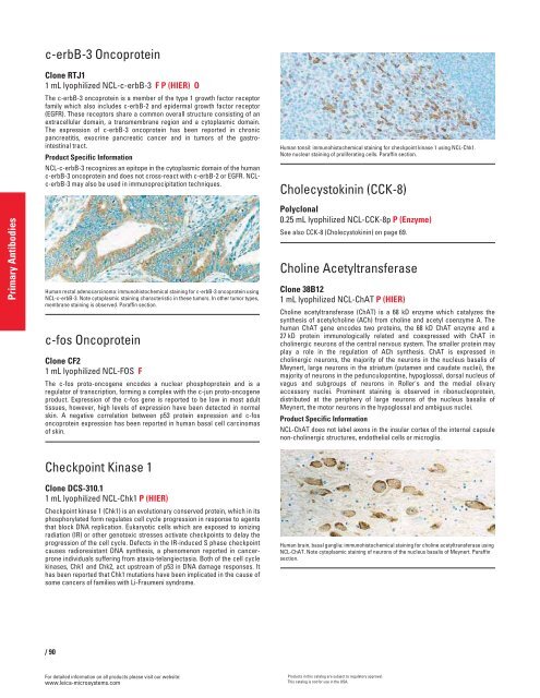
Human tonsil: immunohistochemical staining for checkpoint kinase 1 using NCL-Chk1.<br />
Note nuclear staining of proliferating cells. Paraffin section.<br />
Cholecystokinin (CCK-8)<br />
Polyclonal<br />
0.25 mL lyophilized NCL-CCK-8p P (Enzyme)<br />
See also CCK-8 (Cholecystokinin) on page 69.<br />
Choline Acetyltransferase<br />
Clone 38B12<br />
1 mL lyophilized NCL-ChAT P (HIER)<br />
Choline acetyltransferase (ChAT) is a 68 kD enzyme which catalyzes the<br />
synthesis of acetylcholine (ACh) from choline and acetyl coenzyme A. The<br />
human ChAT gene encodes two proteins, the 68 kD ChAT enzyme and a<br />
27 kD protein immunologically related and coexpressed with ChAT in<br />
cholinergic neurons of the central nervous system. The smaller protein may<br />
play a role in the regulation of ACh synthesis. ChAT is expressed in<br />
cholinergic neurons, the majority of the neurons in the nucleus basalis of<br />
Meynert, large neurons in the striatum (putamen and caudate nuclei), the<br />
majority of neurons in the pedunculopontine, hypoglossal, dorsal nucleus of<br />
vagus and subgroups of neurons in Roller's and the medial olivary<br />
accessory nuclei. Prominent staining is observed in ribonucleoprotein,<br />
distributed at the periphery of large neurons of the nucleus basalis of<br />
Meynert, the motor neurons in the hypoglossal and ambiguus nuclei.<br />
Product Specific Information<br />
NCL-ChAT does not label axons in the insular cortex of the internal capsule<br />
non-cholinergic structures, endothelial cells or microglia.<br />
Human brain, basal ganglia: immunohistochemical staining for choline acetyltransferase using<br />
NCL-ChAT. Note cytoplasmic staining of neurons of the nucleus basalis of Meynert. Paraffin<br />
section.<br />
Products in this catalog are subject to regulatory approval.<br />
This catalog is not for use in the USA.


