

Labelling Review row-Online
Labelling Review row-Online
Labelling Review row-Online

Create successful ePaper yourself
Turn your PDF publications into a flip-book with our unique Google optimized e-Paper software.
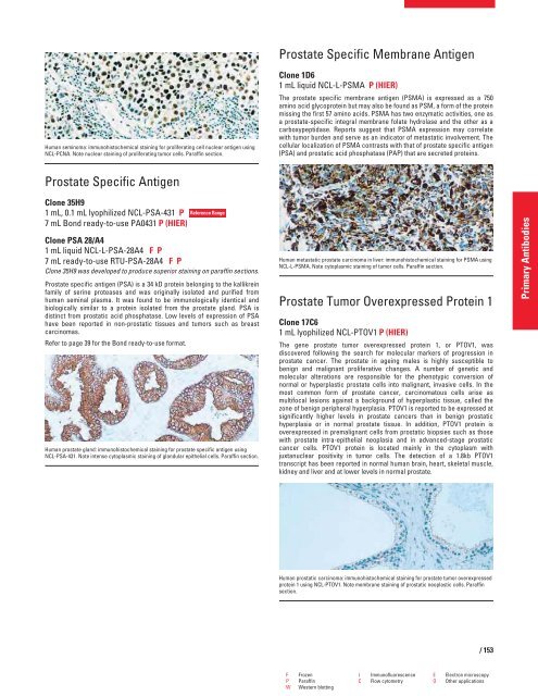
Human seminoma: immunohistochemical staining for proliferating cell nuclear antigen using<br />
NCL-PCNA. Note nuclear staining of proliferating tumor cells. Paraffin section.<br />
Prostate Specific Antigen<br />
Clone 35H9<br />
1 mL, 0.1 mL lyophilized NCL-PSA-431 P<br />
7 mL Bond ready-to-use PA0431 P (HIER)<br />
Reference Range<br />
Clone PSA 28/A4<br />
1 mL liquid NCL-L-PSA-28A4 FP<br />
7 mL ready-to-use RTU-PSA-28A4 FP<br />
Clone 35H9 was developed to produce superior staining on paraffin sections.<br />
Prostate specific antigen (PSA) is a 34 kD protein belonging to the kallikrein<br />
family of serine proteases and was originally isolated and purified from<br />
human seminal plasma. It was found to be immunologically identical and<br />
biologically similar to a protein isolated from the prostate gland. PSA is<br />
distinct from prostatic acid phosphatase. Low levels of expression of PSA<br />
have been reported in non-prostatic tissues and tumors such as breast<br />
carcinomas.<br />
Refer to page 39 for the Bond ready-to-use format.<br />
Human prostate gland: immunohistochemical staining for prostate specific antigen using<br />
NCL-PSA-431. Note intense cytoplasmic staining of glandular epithelial cells. Paraffin section.<br />
Prostate Specific Membrane Antigen<br />
Clone 1D6<br />
1 mL liquid NCL-L-PSMA P (HIER)<br />
The prostate specific membrane antigen (PSMA) is expressed as a 750<br />
amino acid glycoprotein but may also be found as PSM, a form of the protein<br />
missing the first 57 amino acids. PSMA has two enzymatic activities, one as<br />
a prostate-specific integral membrane folate hydrolase and the other as a<br />
carboxypeptidase. Reports suggest that PSMA expression may correlate<br />
with tumor burden and serve as an indicator of metastatic involvement. The<br />
cellular localization of PSMA contrasts with that of prostate specific antigen<br />
(PSA) and prostatic acid phosphatase (PAP) that are secreted proteins.<br />
Human metastatic prostate carcinoma in liver: immunohistochemical staining for PSMA using<br />
NCL-L-PSMA. Note cytoplasmic staining of tumor cells. Paraffin section.<br />
Prostate Tumor Overexpressed Protein 1<br />
Clone 17C6<br />
1 mL lyophilized NCL-PTOV1 P (HIER)<br />
The gene prostate tumor overexpressed protein 1, or PTOV1, was<br />
discovered following the search for molecular markers of progression in<br />
prostate cancer. The prostate in ageing males is highly susceptible to<br />
benign and malignant proliferative changes. A number of genetic and<br />
molecular alterations are responsible for the phenotypic conversion of<br />
normal or hyperplastic prostate cells into malignant, invasive cells. In the<br />
most common form of prostate cancer, carcinomatous cells arise as<br />
multifocal lesions against a background of hyperplastic tissue, called the<br />
zone of benign peripheral hyperplasia. PTOV1 is reported to be expressed at<br />
significantly higher levels in prostate cancers than in benign prostatic<br />
hyperplasia or in normal prostate tissue. In addition, PTOV1 protein is<br />
overexpressed in premalignant cells from prostatic biopsies such as those<br />
with prostate intra-epithelial neoplasia and in advanced-stage prostatic<br />
cancer cells. PTOV1 protein is located mainly in the cytoplasm with<br />
juxtanuclear positivity in tumor cells. The detection of a 1.8kb PTOV1<br />
transcript has been reported in normal human brain, heart, skeletal muscle,<br />
kidney and liver and at lower levels in normal prostate.<br />
Human prostatic carcinoma: immunohistochemical staining for prostate tumor overexpressed<br />
protein 1 using NCL-PTOV1. Note membrane staining of prostatic neoplastic cells. Paraffin<br />
section.<br />
F Frozen I Immunofluorescence E Electron microscopy<br />
P Paraffin C Flow cytometry O Other applications<br />
W Western blotting<br />
/ 153<br />
Primary Antibodies


