Richtlijn: Mammacarcinoom (2.0) - Kwaliteitskoepel
Richtlijn: Mammacarcinoom (2.0) - Kwaliteitskoepel
Richtlijn: Mammacarcinoom (2.0) - Kwaliteitskoepel

Create successful ePaper yourself
Turn your PDF publications into a flip-book with our unique Google optimized e-Paper software.
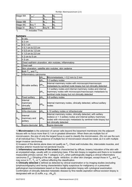
Stage IIIA T 0-2 * N 2 M 0<br />
T 3 N 1-2 M 0<br />
Stage IIIB T 4 N 0-2 M 0<br />
Stage IIIC Any T N 3 M 0<br />
Stage IV Any T Any N M 1<br />
*) T1 includes T1mi.<br />
<strong>Richtlijn</strong>: <strong>Mammacarcinoom</strong> (<strong>2.0</strong>)<br />
Summary<br />
Tis In situ<br />
T1 ≤ 2 cm<br />
T1mi ≤ 0,1 cm<br />
T1a > 0,1 cm to 0,5 cm<br />
T1b > 0,5 cm to 1,0 cm<br />
T1c > 1,0 cm to 2,0 cm<br />
T2 > 2 cm to 5 cm<br />
T3 > 5 cm<br />
T4 Chest wall/skin ulceration, skin nodules, inflammatory<br />
T4a Chest wall<br />
T4b Skin ulceration, satellite skin nodules, skin oedema<br />
T4c Both T4a and T4b T4d Inflammatory carcinoma<br />
pN1mi Micrometastasis, > 0,2 mm to 2 mm<br />
pN1a 1-3 axillary nodes<br />
N1 Movable axillary pN1b Internal mammary nodes with microscopic/macroscopic<br />
metastasis by sentinel node biopsy but not clinically detected<br />
1-3 axillary nodes and internal mammary nodes and internal<br />
pN1c mammary nodes with microscopic/macroscopic metastasis by<br />
sentinel node biopsy but not clinically detected<br />
N2a Fixed axillary<br />
Internal<br />
pN2a 4-9 axillary nodes<br />
N2b mammary<br />
clinically<br />
apparent<br />
pN2b Internal mammary nodes, clinically detected, without axillary<br />
nodes<br />
N3a Infra-clavicular pN3a ≥ 10 axillary nodes or infraclavicular<br />
N3b Internal<br />
mammary and<br />
axillary<br />
pN3b Internal mammary nodes, clinically detected, with axillary<br />
node(s) or > 3 axillary nodes and internal axillary mammary<br />
nodes with microscopic metastasis by sentinal node biopsy but<br />
not clinically detected<br />
N3c Supra-clavicular pN3c Supra-clavicular<br />
1) Microinvasion is the extension of cancer cells beyond the basement membrane into the adjacent<br />
tissues with no focus more than 0,1 cm in greatest dimension. When there are multiple foci of<br />
microinvasion, the size of only the largest focus is used to classify the microinvasion. (Do not use the sum<br />
of all individual foci.) The presence of multiple foci of microinvasion should be noted, as it is with multiple<br />
larger invasive carcinomas.<br />
2) Invasion of the dermis alone does not qualify as T 4 . Chest wall includes ribs, intercostals muscles, and<br />
serratus anterior muscle but not perctoral muscle.<br />
3) Inflammatory carcinoma of the breast is characterized by diffuse, brawny induration of the skin with<br />
an erysipeloid edge, usually with no underlying mass. If the skin biopsy is negative and there is no localized<br />
measurable primary cancer, the T category is pT X when pathologically staging a clinical inflammatory<br />
carcinoma (T 4d ). Dimpling of the skin, nipple retraction, or other skin changes, except those in T 4b and T 4d ,<br />
may occur in T 1 , T 2 , or T 3 without affecting the classification.<br />
4) Clinically detected is defined as detected by clinical examination or by imaging studies (excluding<br />
lymphoscintigraphy) and having characteristics highly suspicious for malignancy or a presumed<br />
pathological macrometastasis based on fine-needle aspiration biopsy with cytological examination.<br />
Confirmation of clinically detected metastatic disease by fine-needle aspiration without excision biopsy is<br />
designated with an (f) suffix, e.g., cN 3a (f).<br />
03/27/2012 <strong>Mammacarcinoom</strong> (<strong>2.0</strong>) 198