Attention! Your ePaper is waiting for publication!
By publishing your document, the content will be optimally indexed by Google via AI and sorted into the right category for over 500 million ePaper readers on YUMPU.
This will ensure high visibility and many readers!

Your ePaper is now published and live on YUMPU!
You can find your publication here:
Share your interactive ePaper on all platforms and on your website with our embed function

Sorted By Test Name - Mayo Medical Laboratories
Sorted By Test Name - Mayo Medical Laboratories
Sorted By Test Name - Mayo Medical Laboratories
You also want an ePaper? Increase the reach of your titles
YUMPU automatically turns print PDFs into web optimized ePapers that Google loves.
MPSU<br />
8823<br />
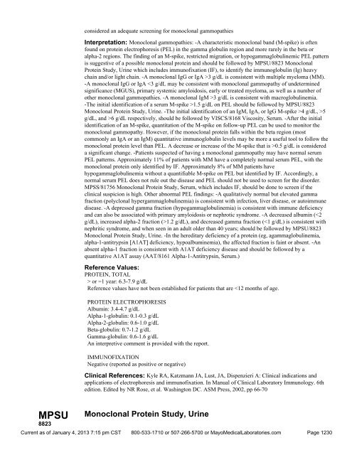
considered an adequate screening for monoclonal gammopathies<br />
Interpretation: Monoclonal gammopathies: -A characteristic monoclonal band (M-spike) is often<br />
found on protein electrophoresis (PEL) in the gamma globulin region and more rarely in the beta or<br />
alpha-2 regions. The finding of an M-spike, restricted migration, or hypogammaglobulinemic PEL pattern<br />
is suggestive of a possible monoclonal protein and should be followed by MPSU/8823 Monoclonal<br />
Protein Study, Urine which includes immunofixation (IF), to identify the immunoglobulin (Ig) heavy<br />
chain and/or light chain. -A monoclonal IgG or IgA >3 g/dL is consistent with multiple myeloma (MM).<br />
-A monoclonal IgG or IgA 3 g/dL is consistent with macroglobulinemia.<br />
-The initial identification of a serum M-spike >1.5 g/dL on PEL should be followed by MPSU/8823<br />
Monoclonal Protein Study, Urine. -The initial identification of an IgM, IgA, or IgG M-spike >4 g/dL, >5<br />
g/dL, and >6 g/dL respectively, should be followed by VISCS/8168 Viscosity, Serum. -After the initial<br />
identification of an M-spike, quantitation of the M-spike on follow-up PEL can be used to monitor the<br />
monoclonal gammopathy. However, if the monoclonal protein falls within the beta region (most<br />
commonly an IgA or an IgM) quantitative immunoglobulin levels may be more a useful tool to follow the<br />
monoclonal protein level than PEL. A decrease or increase of the M-spike that is >0.5 g/dL is considered<br />
a significant change. -Patients suspected of having a monoclonal gammopathy may have normal serum<br />
PEL patterns. Approximately 11% of patients with MM have a completely normal serum PEL, with the<br />
monoclonal protein only identified by IF. Approximately 8% of MM patients have<br />
hypogammaglobulinemia without a quantifiable M-spike on PEL but identified by IF. Accordingly, a<br />
normal serum PEL does not rule out the disease and PEL should not be used to screen for the disorder.<br />
MPSS/81756 Monoclonal Protein Study, Serum, which includes IF, should be done to screen if the<br />
clinical suspicion is high. Other abnormal PEL findings: -A qualitatively normal but elevated gamma<br />
fraction (polyclonal hypergammaglobulinemia) is consistent with infection, liver disease, or autoimmune<br />
disease. -A depressed gamma fraction (hypogammaglobulinemia) is consistent with immune deficiency<br />
and can also be associated with primary amyloidosis or nephrotic syndrome. -A decreased albumin (1.2 g/dL), and decreased gamma fraction ( or =1 year: 6.3-7.9 g/dL<br />
Reference values have not been established for patients that are
RMPSU 60069 MPSS 81756 Monoclonal Protein Study, Random, Urine Clinical Information: Urine proteins can be grouped into 5 fractions by protein electrophoresis: -Albumin -Alpha-1 -Alpha-2 -Beta-globulin -Gamma globulin The urine total protein concentration, the electrophoretic pattern, and the presence of a monoclonal immunoglobulin light chain may be characteristic of monoclonal gammopathies such as multiple myeloma, primary systemic amyloidosis, and light-chain deposition disease. The use of a random urine specimen is sufficient for identifying the presence or absence of a monoclonal immunoglobulin, but a 24 hour specimen is preferred for quantitating and monitoring the abnormality. See An Expanded Algorithm for the Laboratory Evaluation of Suspected Multiple Myeloma in Special Instructions and the Laboratory Approach to the Diagnosis of Amyloidosis algorithm in Special Instructions. Also see Diagnosis and Monitoring of Multiple Myeloma in Publications. Useful For: Diagnosing monoclonal gammopathies Interpretation: A characteristic monoclonal band (M-spike) is often found in the urine of patients with monoclonal gammopathies. The initial identification of an M-spike or an area of restricted migration is followed by immunofixation to identify the immunoglobulin heavy chain and/or light chain. Immunoglobulin free light chains as well as heavy chain fragments may be seen in the urine of patients with monoclonal gammopathies. The presence of a monoclonal light-chain M-spike of >1 g/24 hours is consistent with a diagnosis of multiple myeloma or macroglobulinemia. The presence of a small amount of monoclonal light chain and proteinuria (total protein >3 g/24 hrs) that is predominantly albumin is consistent with primary systemic amyloidosis (AL) or light-chain deposition disease (LCDD). Because patients with AL or LCDD may have elevated urinary protein without an identifiable M-spike, urine protein electrophoresis is not considered an adequate screen for these disorders and immunofixation is also performed. Reference Values: ELECTROPHORESIS, PROTEIN The following fractions, if present, will be reported as a percent of the total protein: Albumin Alpha-1-globulin Alpha-2-globulin Beta-globulin Gamma-globulin No reference values apply to random urine. Clinical References: Kyle RA, Katzmann JA, Lust JA, Dispenzieri A: Clinical indications and applications of electrophoresis and immunofixation. In Manual of Clinical Laboratory Immunology. 6th edition. Edited by NR Rose, et al. Washington, DC, ASM Press, 2002, pp 66-67 Monoclonal Protein Study, Serum Clinical Information: Serum proteins can be grouped into 5 fractions by protein electrophoresis: -Albumin, which represents almost 2/3 of the total serum protein -Alpha-1, composed primarily of alpha-1-antitrypsin (A1AT), an alpha-1-acid glycoprotein -Alpha-2, composed primarily of alpha-2-macroglobulin and haptoglobin -Beta, composed primarily of transferrin and complement C3 -Gamma, composed primarily of immunoglobulins (Ig) The concentration of these fractions and the electrophoretic pattern may be characteristic of diseases such as monoclonal gammopathies, A1AT deficiency disease, nephrotic syndrome, and inflammatory processes associated with infection, liver disease, and autoimmune diseases. See Laboratory Screening <strong>Test</strong>s for Suspected Multiple Myeloma and the Laboratory Approach to the Diagnosis of Amyloidosis algorithm in Special Instructions. Also see Diagnosis and Monitoring of Multiple Myeloma in Publications. Useful For: Diagnosis of monoclonal gammopathies, when used in conjunction with urine monoclonal studies Monitoring patients with monoclonal gammopathies Protein electrophoresis alone is not Current as of January 4, 2013 7:15 pm CST 800-533-1710 or 507-266-5700 or <strong>Mayo</strong><strong>Medical</strong><strong>Laboratories</strong>.com Page 1229
MPSU 8823 considered an adequate screening for monoclonal gammopathies Interpretation: Monoclonal gammopathies: -A characteristic monoclonal band (M-spike) is often found on protein electrophoresis (PEL) in the gamma globulin region and more rarely in the beta or alpha-2 regions. The finding of an M-spike, restricted migration, or hypogammaglobulinemic PEL pattern is suggestive of a possible monoclonal protein and should be followed by MPSU/8823 Monoclonal Protein Study, Urine which includes immunofixation (IF), to identify the immunoglobulin (Ig) heavy chain and/or light chain. -A monoclonal IgG or IgA >3 g/dL is consistent with multiple myeloma (MM). -A monoclonal IgG or IgA 3 g/dL is consistent with macroglobulinemia. -The initial identification of a serum M-spike >1.5 g/dL on PEL should be followed by MPSU/8823 Monoclonal Protein Study, Urine. -The initial identification of an IgM, IgA, or IgG M-spike >4 g/dL, >5 g/dL, and >6 g/dL respectively, should be followed by VISCS/8168 Viscosity, Serum. -After the initial identification of an M-spike, quantitation of the M-spike on follow-up PEL can be used to monitor the monoclonal gammopathy. However, if the monoclonal protein falls within the beta region (most commonly an IgA or an IgM) quantitative immunoglobulin levels may be more a useful tool to follow the monoclonal protein level than PEL. A decrease or increase of the M-spike that is >0.5 g/dL is considered a significant change. -Patients suspected of having a monoclonal gammopathy may have normal serum PEL patterns. Approximately 11% of patients with MM have a completely normal serum PEL, with the monoclonal protein only identified by IF. Approximately 8% of MM patients have hypogammaglobulinemia without a quantifiable M-spike on PEL but identified by IF. Accordingly, a normal serum PEL does not rule out the disease and PEL should not be used to screen for the disorder. MPSS/81756 Monoclonal Protein Study, Serum, which includes IF, should be done to screen if the clinical suspicion is high. Other abnormal PEL findings: -A qualitatively normal but elevated gamma fraction (polyclonal hypergammaglobulinemia) is consistent with infection, liver disease, or autoimmune disease. -A depressed gamma fraction (hypogammaglobulinemia) is consistent with immune deficiency and can also be associated with primary amyloidosis or nephrotic syndrome. -A decreased albumin (1.2 g/dL), and decreased gamma fraction ( or =1 year: 6.3-7.9 g/dL Reference values have not been established for patients that are
- Page 1 and 2:
Rochester 2013 Interpretive Handboo
- Page 3 and 4:
Policies - Mayo Medical Laboratorie
- Page 5 and 6:
Policies - Mayo Medical Laboratorie
- Page 7 and 8:
Policies - Mayo Medical Laboratorie
- Page 9 and 10:
Policies - Mayo Medical Laboratorie
- Page 11 and 12:
Policies - Mayo Medical Laboratorie
- Page 13 and 14:
TTIG 82506 DHVD 8822 Tetanus Toxoid
- Page 15 and 16:
DCRN 8847 Identification of patient
- Page 17 and 18:
DOC 8547 function primarily, 18-hyd
- Page 19 and 20:
FDSOX 91690 THCM 84284 11-Desoxycor
- Page 21 and 22:
FBP1 86208 Normal: or =1.5 ng/mL;
- Page 23 and 24:
17OHP 81151 Pediatric Reference Ran
- Page 25 and 26:
OHPG 9231 2000;52(5):601-607 4. Kao
- Page 27 and 28:
FP73 88541 Interpretation: The pres
- Page 29 and 30:
OH21 81970 21-deoxycortisol may be
- Page 31 and 32:
CYPKP 89082 transcriptionally activ
- Page 33 and 34:
25HDN 83670 25-Hydroxyvitamin D2 an
- Page 35 and 36:
F5HAR 57333 HIAA 9248 can also be a
- Page 37 and 38:
6MAMU 89605 converts heroin into 6-
- Page 39 and 40:
ACAC 82757 ACANT 80401 increase the
- Page 41 and 42:
ACM 8698 FACTO 90247 1 0.35-0.69 Eq
- Page 43 and 44:
ACHS 8522 Clinical Information: Neu
- Page 45 and 46:
ACT 8221 APT 9058 and isolation of
- Page 47 and 48:
AHPS 9022 ancestry. Homozygosity fo
- Page 49 and 50:
FAML secretions, but it is not comm
- Page 51 and 52:
ACRN 82413 Acylcarnitines, Quantita
- Page 53 and 54:
or =8 years:
- Page 55 and 56:
ADM13 61212 studies (enzyme assay,
- Page 57 and 58:
FADA 91554 81444 (50-70 x normal) A
- Page 59 and 60:
FADE 91670 LADV 89074 LCADP 89887 A
- Page 61 and 62:
FADMK 91925 RACTH 82140 ADmark Phos
- Page 63 and 64:
AGXMS 89915 7 year 2-88 8 year 5-71
- Page 65 and 66:
the third decade of life, but can o
- Page 67 and 68:
FALUF 57286 ALB 8436 involved in th
- Page 69 and 70:
FALCO 90084 ALS 8363 ALDNA 15150 th
- Page 71 and 72:
ALDU 8556 regulator of the synthesi
- Page 73 and 74:
ALKI 89503 11 years: 185-507 U/L 12
- Page 75 and 76:
17-23 years: 52-144 U/L 24-45 years
- Page 77 and 78:
FALMD 92001 ALM 82882 Almond (Amygd
- Page 79 and 80:
FASU 91221 FA1AG 90464 19-70+ years
- Page 81 and 82:
A1APP 26953 genotype is at greater
- Page 83 and 84:
A1M24 81036 RA1M 84448 605-608 2. M
- Page 85 and 86:
A2M 9270 AAMY 82866 Alpha-2-Macrogl
- Page 87 and 88:
AFP 8162 hepatocellular carcinoma,
- Page 89 and 90:
AFPSF 8876 Screen [Second Trimester
- Page 91 and 92:
FUCW 8814 molecules in the tissues
- Page 93 and 94:
AGA 8785 disease. In female patient
- Page 95 and 96:
AGPB 9499 IDSWB 60618 disease. In T
- Page 97 and 98:
IDST 8780 categorized into 3 syndro
- Page 99 and 100:
MANT 8773 MANN 8772 5 50.0-99.9 Str
- Page 101 and 102:
ANAS 8782 APGH 9003 Alpha-N-Acetylg
- Page 103 and 104:
ALPA 500155 Tanner II-IV*: < or =1.
- Page 105 and 106:
ALUR 86377 -Aluminum-laden albumin
- Page 107 and 108:
86375 exposure. This was done by sw
- Page 109 and 110:
FAMAN 91132 AMKPK 82112 possible ev
- Page 111 and 112:
AAMSD 60200 susceptible strains of
- Page 113 and 114:
Serine (Ser) 69-271 71-208 63-187 H
- Page 115 and 116:
Glutamine Gln 139-2985 263 -2979 15
- Page 117 and 118:
AAUCD 60202 Glutamic Acid (Glu) 1-M
- Page 119 and 120:
FALAU 57350 ALADW to secondary inhi
- Page 121 and 122:
AMIO 9247 dehydratase (ALAD) activi
- Page 123 and 124:
AMOBS 8325 FAMOX 80450 FMAXN 57245
- Page 125 and 126:
AMPCC 61514 receptors that mediate
- Page 127 and 128:
FAMPH 90113 AMPHU 8257 MJ, Parks PM
- Page 129 and 130:
AMBF 8371 immune response to allerg
- Page 131 and 132:
PAMYB 5079 PAMY 80376 Clinical Refe
- Page 133 and 134:
AMSU 8356 AMS 8352 Amylase, Timed C
- Page 135 and 136:
AMYL 83667 AMYKM 83705 provided. Re
- Page 137 and 138:
ANAP 81157 TTR-associated familial
- Page 139 and 140:
F3AAG 57109 ANST 9709 Class IgE kU/
- Page 141 and 142:
FPOC 81081 correlate only modestly
- Page 143 and 144:
FANGI 90429 FANG 90428 Interpretati
- Page 145 and 146:
FANSD 91955 FAEAB 91854 immunoglobu
- Page 147 and 148:
FANBF 57173 FCLNE 91321 FPHET 91322
- Page 149 and 150:
ABTIH 9000 ENAE 89035 Antibody Tite
- Page 151 and 152:
MMLYP 81602 MMLRG 81601 Test Perfor
- Page 153 and 154:
MMLSA 56031 MACCL 89218 antimicrobi
- Page 155 and 156:
FAMS 91540 Clinical Information: No
- Page 157 and 158:
VASC 83012 evaluation for infertili
- Page 159 and 160:
ANA2 9026 ATTF 9030 Antinuclear Ant
- Page 161 and 162:
ATTI 9031 Antithrombin Antigen, Pla
- Page 163 and 164:
APO1K 60724 APO2S 60725 Shiller SM,
- Page 165 and 166:
APLAB 80318 APLA1 80309 mutation in
- Page 167 and 168:
APOE 80905 expressing more severe d
- Page 169 and 170:
APR 82835 AWNS 87814 6 > or =100 St
- Page 171 and 172:
ARBOP 83267 Reference values apply
- Page 173 and 174:
distinguishable from comparison gro
- Page 175 and 176:
FARI 57112 FARIX 57123 FCGHP 89858
- Page 177 and 178:
ARSAS 61259 ARSAK number variation
- Page 179 and 180:
ASFRU 84679 forms. Concentrations >
- Page 181 and 182:
ASHA 8651 and their partially detox
- Page 183 and 184:
ASCRU 89890 are nontoxic, inorganic
- Page 185 and 186:
ARSAW 8779 Useful For: Detection of
- Page 187 and 188:
ARSB 8151 ASCRI 82764 Roth. McGraw-
- Page 189 and 190:
AJPWO 88887 Clinical References: 1.
- Page 191 and 192:
AST 8360 clinical manifestations. I
- Page 193 and 194:
FASP 91607 ASPBA 61009 antigen in f
- Page 195 and 196:
SASP 9678 ASPG 86324 Aspergillus fu
- Page 197 and 198:
FAPP 91428 Reference Values: or =1
- Page 199 and 200:
APIN 82803 responsible for elicitin
- Page 201 and 202:
AGIDE 89886 < or =0.02 nmol/L NEURO
- Page 203 and 204:
ALDE 84248 anti-neuronal nuclear au
- Page 205 and 206:
FAVI 91509 AVOC 82812 approximately
- Page 207 and 208:
CD40 89009 conventional cytogenetic
- Page 209 and 210:
80027 IABCS 88800 patients with B-c
- Page 211 and 212:
B-cell-depleting immunotherapy Iden
- Page 213 and 214:
functional classification system fo
- Page 215 and 216:
FBAB 91608 BABG 81128 5785 Corporat
- Page 217 and 218:
GEN 8108 SPUT 8095 Bruyn G, Pieniaz
- Page 219 and 220:
CFRC 89653 EPRP 60235 result in int
- Page 221 and 222:
BAHG 82711 BCYP 82722 electrophores
- Page 223 and 224:
BANA 82746 sensitization to particu
- Page 225 and 226:
BRLY 82687 caused by the release of
- Page 227 and 228:
FBART 91439 BART 81575 amplificatio
- Page 229 and 230:
BARTB 89983 BASL 82489 B. bacillifo
- Page 231 and 232:
BADX 89006 Interpretation: Detectio
- Page 233 and 234:
MBCR 80578 helpful for both prognos
- Page 235 and 236:
FBBCK 91664 FBEAN 91646 ASPE/Lumine
- Page 237 and 238:
BECH 82669 of IC2 (LIT1) is hypothe
- Page 239 and 240:
BEEF 82697 BEETS 82618 Beef, IgE Cl
- Page 241 and 242:
FPHEN 91136 FBEN 90294 BENZU 80370
- Page 243 and 244:
BBEET 82838 BERG 82892 Berlin Beetl
- Page 245 and 246:
AB2GP 86180 Negative (reported as p
- Page 247 and 248:
GB2GP 86182 or =15.0 U/mL (positiv
- Page 249 and 250:
BETA2 80351 patients with APS.(4) A
- Page 251 and 252:
B2MU 300243 B2M 9234 Livanainen M,
- Page 253 and 254:
BGAW 60987 Postmenopausal: 104-1,00
- Page 255 and 256:
BGAT 8008 beta-galactosidase defici
- Page 257 and 258:
BGLT 8787 BGL 8788 B disease. Clini
- Page 259 and 260:
BHCG 61718 Sly syndrome is 1 of the
- Page 261 and 262:
BLACT 8118 BLAC Clinical Informatio
- Page 263 and 264:
and the determination of bicarbonat
- Page 265 and 266:
84357 19701 Interpretation: Total b
- Page 267 and 268:
BFBL 34621 the color of bilirubin a
- Page 269 and 270:
BILIT 81785 Bilirubin, Total, Serum
- Page 271 and 272:
BIOTS 88205 levels of biotinidase,
- Page 273 and 274:
FBISU 91142 LCBK 88910 Test Perform
- Page 275 and 276:
QBKU 87859 Useful For: As a prospec
- Page 277 and 278:
SBLAS 86691 antibodies interact wit
- Page 279 and 280:
80326 BDIAL 83094 night sweats. It
- Page 281 and 282:
UEBF 81834 BWOR 82840 urinary tract
- Page 283 and 284:
BLUE 82359 Clinical Information: Cl
- Page 285 and 286:
BHINT 9027 resorption is balanced b
- Page 287 and 288:
BPRP 80910 FBPTS 57290 An interpret
- Page 289 and 290:
BOAC 9723 BOT 82715 Cypress, CA 906
- Page 291 and 292:
89045 proteins) followed by respira
- Page 293 and 294:
BRAZ 82899 MLH1 promoter hypermethy
- Page 295 and 296:
BMNA 81976 FYSTB 91990 BROC 82817 C
- Page 297 and 298:
BRM 8608 BRUGM 89476 0 Negative 1 0
- Page 299 and 300:
BRUTA 8112 brucellosis may include
- Page 301 and 302:
manifests in male children younger
- Page 303 and 304:
clinical phenotype can vary conside
- Page 305 and 306:
BTKK 89306 Useful For: Confirming a
- Page 307 and 308:
BUCW 82727 XLA patients are diagnos
- Page 309 and 310:
BFTH 82779 identify allergens which
- Page 311 and 312:
BUPIS 89548 BUPM 500038 membrane pr
- Page 313 and 314:
FBUS 91115 BUAUC 83188 increase in
- Page 315 and 316:
FCPEP 91270 anti-insulin autoantibo
- Page 317 and 318:
C1ES 8198 Useful For: Assessment of
- Page 319 and 320:
C1QFX 83374 Reference Values: C1Q B
- Page 321 and 322:
C2 81835 receptors. The absence of
- Page 323 and 324:
FC3D 91725 C4U 88829 C4FX 83391 C3d
- Page 325 and 326:
C5FX 83392 C5DCU 88831 1987;76:939-
- Page 327 and 328:
C7FX 81064 complement system is com
- Page 329 and 330:
C9FX 81066 CABB 86327 4. Gaither TA
- Page 331 and 332:
CDOMB 89539 CDOM 80595 Reference Va
- Page 333 and 334:
CDRU 60156 CAFF 8754 years) is the
- Page 335 and 336:
3 10.7-15.4 17-72 4 11.8-16.2 15-11
- Page 337 and 338:
FCAH1 91275 1 - 7m: Levels decrease
- Page 339 and 340:
Males: Levels increase after the fi
- Page 341 and 342:
Units: ug/dL Age Range Premature (2
- Page 343 and 344:
4 10.7-15.6 47-208 5 11.8-18.6 50-2
- Page 345 and 346:
anging from 40 - 200 between 30 and
- Page 347 and 348:
Clinical Information: In the normal
- Page 349 and 350:
CSRMS 83703 hypoparathyroidism, a s
- Page 351 and 352:
CAU 8594 CAF 8379 An interpretive r
- Page 353 and 354:
CAUR 60157 CAS parathyroid hormone-
- Page 355 and 356:
CACRU 89604 important role in blood
- Page 357 and 358:
CAVPC 83900 excretion of calcium is
- Page 359 and 360:
FCALP 91597 FCAMP 91224 CFTH 82778
- Page 361 and 362:
CANW 81780 CA25 9289 1 0.35-0.69 Eq
- Page 363 and 364:
FCANA 57158 testing often depend up
- Page 365 and 366:
FFTH 90479 FCAPR 90062 CWAY 82493 C
- Page 367 and 368:
CAR 8654 C1011 80682 total and free
- Page 369 and 370:
199PC 89508 199PT 61530 Clinical Re
- Page 371 and 372:
CA19 9288 CDG 89891 Carbohydrate An
- Page 373 and 374:
CHOU 9255 COHBB 8649 isoforms (ie,
- Page 375 and 376:
CEAPT 61528 PFCEA 83742 Carcinoembr
- Page 377 and 378:
CEASF 8918 CARD 82491 Carcinoembryo
- Page 379 and 380:
CPTKM 61121 CARN 8802 screening can
- Page 381 and 382:
cycle, or secondary disturbances in
- Page 383 and 384:
CACTK 61195 CAROB 82368 analysis. U
- Page 385 and 386:
FCASE 91647 caused by the release o
- Page 387 and 388:
FCASO 91995 CBN 82770 sensitivity t
- Page 389 and 390:
COMTO 60336 1 0.35-0.69 Equivocal 2
- Page 391 and 392:
CATU 9276 An interpretive report wi
- Page 393 and 394:
neuropathies are characterized by e
- Page 395 and 396:
CBC 9109 Useful For: Testing for Ig
- Page 397 and 398:
15 days-1 month: 31.0-55.0% 2-5 mon
- Page 399 and 400:
CD20B 89584 CD20 on B Cells Clinica
- Page 401 and 402:
3-5 months: 51-77%* 6-11 months: 49
- Page 403 and 404:
55 years: 49-87% % Helper cells (CD
- Page 405 and 406:
GLICP 89369 essential to serially m
- Page 407 and 408:
T- AND B-CELL QUANTITATION BY FLOW
- Page 409 and 410:
GLIC 89317 H/S ratio: > or =1.0 *Sh
- Page 411 and 412:
a more recent thymic ontogeny where
- Page 413 and 414:
CDKKM 60229 Med Genet 1999;36:518-5
- Page 415 and 416:
CELY 82766 CDCOM 89201 Company, 200
- Page 417 and 418:
CDGF 89200 serology does not exclud
- Page 419 and 420:
CELI 88906 range. For these individ
- Page 421 and 422:
5368 CMA 9278 autoantibodies can be
- Page 423 and 424:
CTSA 81979 sensitization to particu
- Page 425 and 426:
CERE 8364 patients with multiple sc
- Page 427 and 428:
8032 80199 2002 April ;287(16):2114
- Page 429 and 430:
CFTRK 88880 deferens or pancreatiti
- Page 431 and 432:
CCHZ 82752 MCHZ 82751 Test Performe
- Page 433 and 434:
CTRE 82607 likelihood of allergic d
- Page 435 and 436:
CHXP 82494 Clinical Information: Im
- Page 437 and 438:
CHCK 82713 CSPR 82351 5 50.0-99.9 S
- Page 439 and 440:
CHILI 82499 and to define the aller
- Page 441 and 442:
CHRGB 83186 CHPA 80411 FCPP 57339 S
- Page 443 and 444:
CGRNA 61553 Clinical References: 1.
- Page 445 and 446:
FCTRC 91659 CDFAW 110502 are requir
- Page 447 and 448:
FCPC 90439 FCPD 91750 FCHH 90111 pr
- Page 449 and 450:
CLFT 60028 CLBF 8470 CLF 8467 as hi
- Page 451 and 452:
CLSF 8218 CLU 8531 FCCK 90162 > or
- Page 453 and 454:
HDCH 8429 CHOL 8320 Serous Body Flu
- Page 455 and 456:
BHSF 8877 FCHAB 91900 serum may inc
- Page 457 and 458:
CR 86153 concentrations in the abse
- Page 459 and 460:
(5-hydroxytryptamine: 5-HT) and pep
- Page 461 and 462:
POC 8887 CVS 80257 An interpretativ
- Page 463 and 464:
CMS 8696 HBL 8537 deletions in 13q1
- Page 465 and 466:
LN 8911 SCE 926 Chromosome Analysis
- Page 467 and 468:
SBK 89338 to summarize here. The re
- Page 469 and 470:
CHSBP 9023 process. A normal karyot
- Page 471 and 472:
FCLL 83089 Negative Interpretation
- Page 473 and 474:
CHYM 82609 the urine (chyluria) is
- Page 475 and 476:
CTCB 89089 CTCC 89162 This test was
- Page 477 and 478:
RCITR 84773 CITR 9329 Reference Val
- Page 479 and 480:
CLAM 82884 proteins) followed by re
- Page 481 and 482:
CLLM 60490 Reference Values: An int
- Page 483 and 484:
CLOS 80424 CDRP 83124 Sedation has
- Page 485 and 486:
FCMVQ 91734 recurrent suicidal beha
- Page 487 and 488:
F_9 9065 FACTV 9054 Adults: 75-145%
- Page 489 and 490:
F7IS 7809 F8A 9070 prolonged prothr
- Page 491 and 492:
FXCH 89042 F10IS 7811 Coagulation F
- Page 493 and 494:
F_12 9069 COU 80083 Basic Principle
- Page 495 and 496:
COS 80084 COBCU 60353 Cobalt, Serum
- Page 497 and 498:
COKEU 9286 fluid.(7) The first evid
- Page 499 and 500:
CCOC 81542 Interpretation: Compleme
- Page 501 and 502:
COCR 82693 clinical manifestations.
- Page 503 and 504:
COD 82889 Q10 87853 5 50.0-99.9 Str
- Page 505 and 506:
CAGG 8992 FFTYC 91496 CTF 80440 Col
- Page 507 and 508:
CVID 87993 testing often depend upo
- Page 509 and 510:
C4 8171 AH50 88676 Clinical Informa
- Page 511 and 512:
FCCEV 57461 FFDMC 91581 FMDMC 91578
- Page 513 and 514:
CAH21 87815 congenital adrenal hype
- Page 515 and 516:
CTDC 83631 Reference Values: An int
- Page 517 and 518:
CURU Interpretation: The constellat
- Page 519 and 520:
CUCRU 60427 CORI can cause hypocupr
- Page 521 and 522:
CRNP 82718 CORN 82705 Corn Pollen,
- Page 523 and 524:
sex steroids. Synthesis proceeds fr
- Page 525 and 526:
FCBG 90148 FCORT 91644 CRANU 82920
- Page 527 and 528:
CORTU 8546 Cortisol, Serum (ug/dL)
- Page 529 and 530:
CORT 8545 measurements, midnight bl
- Page 531 and 532:
COCRU 88903 serum and urine cortiso
- Page 533 and 534:
hydrocortisone) increases and is fi
- Page 535 and 536:
CSED 82804 Class IgE kU/L Interpret
- Page 537 and 538:
FACX 91340 immune response to aller
- Page 539 and 540:
89366 CPOXS 61263 CPM, 12q15, for W
- Page 541 and 542:
CRANB 86307 bronchospasm) in infant
- Page 543 and 544:
CRDPU 88697 6 > or =100 Strongly po
- Page 545 and 546:
CKMB 82429 CK 8336 Creatine Kinase
- Page 547 and 548:
CREAZ 8472 13-36 months: 0.1-0.4 mg
- Page 549 and 550:
CRBF 8037 RCTUR 83802 guidelines fo
- Page 551 and 552:
CRGSP 83659 CRY_S 80988 Clinical Re
- Page 553 and 554:
CCRYP 86166 CRYPF 60320 in serum sp
- Page 555 and 556:
CRYPU 60319 Clinical Information: C
- Page 557 and 558:
CUKE 82861 Reference Values: Anti-T
- Page 559 and 560:
WHTC 82915 proteins) followed by re
- Page 561 and 562:
VRID2 5190 FCMSD 92004 CURR 82498 C
- Page 563 and 564:
60506 CIFS 8052 Cutaneous Anaplasti
- Page 565 and 566:
CYAN 8691 GRP 8771 pattern at the B
- Page 567 and 568:
CYCL 81506 CYCSP 8931 Reference Ran
- Page 569 and 570:
insufficiency. The incidence of CF
- Page 571 and 572:
CYSR 81067 distinguished by the pat
- Page 573 and 574:
drugs and environmental factors. Us
- Page 575 and 576:
2C19S 60439 activity include: -Broc
- Page 577 and 578:
including the intestines and liver.
- Page 579 and 580:
2C9SO 60337 Interpretation: An inte
- Page 581 and 582:
2613delAGA Decreased activity *10 1
- Page 583 and 584:
2D6TO 60340 an interpretation indic
- Page 585 and 586:
antidepressants, antiemetics, antih
- Page 587 and 588:
3A4O 61242 individual is homozygous
- Page 589 and 590:
CMG 80750 known. A ratio of > or =2
- Page 591 and 592:
e seen after acute illness, immunos
- Page 593 and 594:
and have cytotoxic function in resp
- Page 595 and 596:
ANCA 9441 Clinical Information: Cyt
- Page 597 and 598:
DLAC 8878 DLAU 8873 fibrinogen equi
- Page 599 and 600:
DAND 82694 FDANT 90363 5 50.0-99.9
- Page 601 and 602:
DATRE 82481 9803 DEEP 82144 Date, T
- Page 603 and 604:
observed at birth. Levels then decl
- Page 605 and 606:
diagnostic accuracy than DHEA-S mea
- Page 607 and 608:
FDOC 90134 5468 children. Character
- Page 609 and 610:
DESIP 81854 DSG13 83680 1 0.35-0.69
- Page 611 and 612:
FDXM 91956 FDEXA 91777 diagnosis, t
- Page 613 and 614:
DIA 8629 FDIGS 91454 Clinical Refer
- Page 615 and 616:
DIG 8674 FDPD 57141 American Associ
- Page 617 and 618:
DILL 82602 serum levels. Patients w
- Page 619 and 620:
DIP 83262 DTAB 83269 FDIPY 90371 Co
- Page 621 and 622:
FDISP 91595 DSP 8220 Sucrase: Range
- Page 623 and 624:
FDKYL 91960 DOGD 60108 Increases of
- Page 625 and 626:
DRD3O 60342 should be monitored clo
- Page 627 and 628:
DRD4O 60344 Reference Values: An in
- Page 629 and 630:
FDOXY 90061 CDAU1 80917 doxepin and
- Page 631 and 632:
CDAU3 80919 spectrometry (GC-MS) th
- Page 633 and 634:
IDOAU 8248 (GC-FID) the following d
- Page 635 and 636:
DABAR 505335 Urine Clinical Informa
- Page 637 and 638:
DMETH 505343 DPCP 505339 EMIT cutof
- Page 639 and 640:
DTHC 505331 and kidney. Toxic manif
- Page 641 and 642:
DSS 8421 CDAS 500752 Drugs of toxic
- Page 643 and 644:
FBDAS 91776 FMP10 57505 FBD 57117 D
- Page 645 and 646:
DASM4 60553 spectrometry (LC-MS/MS)
- Page 647 and 648:
DULOX 89305 immunoglobulin E (IgE)-
- Page 649 and 650:
ESYC 82721 infected individuals, it
- Page 651 and 652:
FECHC 91342 ECHO 80293 concentratio
- Page 653 and 654:
FEGFR 91903 FEGGW 91976 EGG 82872 W
- Page 655 and 656:
EGGP 82477 concentration of IgE ant
- Page 657 and 658:
FECHA 91710 EHRL 84319
- Page 659 and 660:
ELDR 82392 ELPN 87972 No pediatric
- Page 661 and 662:
EFP 81488 SODIUM 0-15 years: not es
- Page 663 and 664:
PEL 80085 chain fragments as well a
- Page 665 and 666:
ELM 82672 PROTEIN, TOTAL > or =18 y
- Page 667 and 668:
Reference Range: IgG
- Page 669 and 670:
5362 FEMA 91836 EMA 9360 anatomic p
- Page 671 and 672:
SAM 9049 FEHA 91932 Interpretation:
- Page 673 and 674:
FENTQ 91312 LENT 80066 INTERPRETIVE
- Page 675 and 676:
FEPHD 90109 EPUR 82854 presence of
- Page 677 and 678:
SEBV 84421 Clinical Information: Cl
- Page 679 and 680:
80786 EBVA 8891 antigen. The presen
- Page 681 and 682:
LEBV 81239 EBVB 87439 with type 1 N
- Page 683 and 684:
REVP 84160 M, Emre S, et al: Prospe
- Page 685 and 686:
EPOR 61679 FESC 91458 FFES no eryth
- Page 687 and 688:
primarily in ovaries and testes by
- Page 689 and 690:
UE3 81711 Stage V 18 years 10-40 pg
- Page 691 and 692:
ESTF 84230 Immunohistochemistry, Ma
- Page 693 and 694:
preparations). The gonadotrophin-re
- Page 695 and 696:
E1 81418 Stage V 14.5 years 15-350
- Page 697 and 698:
are due to problems in androgen sig
- Page 699 and 700:
ETOHU 500323 ETX 8769 Interpretatio
- Page 701 and 702:
EOXD 82767 EUCL 82758 Ethylene Oxid
- Page 703 and 704:
EHOR 82662 0 Negative 1 0.35-0.69 E
- Page 705 and 706:
83363 fluorescence in situ hybridiz
- Page 707 and 708:
F9INH 83103 lysosomes of both perip
- Page 709 and 710:
F8INH 83102 FX13M 57302 FOGT Clinic
- Page 711 and 712:
FAPKM 83001 and to define the aller
- Page 713 and 714:
FD 85319 LDLRS 81013 features This
- Page 715 and 716:
LDLM 89073 Useful For: Genetic test
- Page 717 and 718:
FANCA 85318 FATF 8310 separately or
- Page 719 and 720:
FAPCP 82042 Clinic or elsewhere, an
- Page 721 and 722:
or =18 years: 30-450 nmol/mL Hexade
- Page 723 and 724:
Hexacosanoic Acid, C26:0 0.00-1.30
- Page 725 and 726:
1-17 years: 9-130 nmol/mL > or =18
- Page 727 and 728:
FAPM 81939 or =18 years: 7.3-16.8
- Page 729 and 730:
POX 81369 oxidation disorders. Ann
- Page 731 and 732:
FBN1 89308 range of variability, in
- Page 733 and 734:
FETH2 81880 FFAPL 57379 Delineation
- Page 735 and 736:
FOBT 60693 Useful For: Suggesting p
- Page 737 and 738:
FENTU 89655 testing often depend up
- Page 739 and 740:
FERR 8689 Clinical Information: Cli
- Page 741 and 742:
FECHK 60372 biochemical genetic tes
- Page 743 and 744:
FMBNY 30320 pregnancy, as well as b
- Page 745 and 746:
FGAKM 60722 in order to provide an
- Page 747 and 748:
FIBR 8482 FBC 80333 FGF23 88662 Fem
- Page 749 and 750:
FFIL4 90068 FIL 9232 The detected c
- Page 751 and 752:
FANT 82698 FBSH 82735 Laboratory Me
- Page 753 and 754:
9804 FLEC 9243 growth factor-bindin
- Page 755 and 756:
FFLRO 91795 FL 8641 impaired patien
- Page 757 and 758:
FFLUR 90091 17BFP 89739 ST. PAUL, M
- Page 759 and 760:
FSH 8670 folate deficiency states.
- Page 761 and 762:
FDP1 86207 antibodies interact with
- Page 763 and 764:
FOOD4 81872 3 3.50-17.4 Positive 4
- Page 765 and 766:
FOOD1 81868 sensitivity to inhalant
- Page 767 and 768:
FOOD3 81871 FRMH 82869 Clinical Ref
- Page 769 and 770:
9881 60694 Reporting limit determin
- Page 771 and 772:
FRANC 91552 other FMR1-related diso
- Page 773 and 774:
FFRED 91819 FFRBS 60476 Reference V
- Page 775 and 776:
FRUCT 81610 FROS 81164 GFDMS protei
- Page 777 and 778:
FSS 83121 FSC 83120 family member.
- Page 779 and 780:
FGEN 84389 FVAG 5184 after 30 days
- Page 781 and 782:
FFURO 91119 FUSM 82750 Furosemide (
- Page 783 and 784:
GABA 80826 cerebrospinal fluid may
- Page 785 and 786:
GDUR 89316 myocardium have also bee
- Page 787 and 788:
GALK 8628 associated with the nephr
- Page 789 and 790:
GALU 8765 GAL1P 80337 Galactose, Qu
- Page 791 and 792:
GALTP 80341 of GALT enzyme activity
- Page 793 and 794:
GALTK 84367 (galactose and galactos
- Page 795 and 796:
CBGT 8297 frequently observed mutat
- Page 797 and 798:
GCC 81981 Galectin-3 is a biomarker
- Page 799 and 800:
GGT 8677 interpretation and reporti
- Page 801 and 802:
GM1B 83189 Ganglioside Antibody Pan
- Page 803 and 804:
FGIP 90171 GAST 8512 1 0.35-0.69 Eq
- Page 805 and 806:
GBAMS 60711 GBAKM 60712 fasting per
- Page 807 and 808:
GELA 86326 diagnosis in at-risk pre
- Page 809 and 810:
GENPK 84695 GENT 81750 amyloidosis,
- Page 811 and 812:
GERB 82545 89713 Am Fam Physician 1
- Page 813 and 814:
GRAB 80628 GIAR 80231 3 3.50-17.4 P
- Page 815 and 816:
DGLDN 89031 2 0.70-3.49 Positive 3
- Page 817 and 818:
equires a jejunal biopsy demonstrat
- Page 819 and 820:
FGLIP 91097 GBM 8106 Disease Diagno
- Page 821 and 822:
GPI 9158 GLBF 8343 then rise again
- Page 823 and 824:
RGLUR 89847 GLSF 152 GLUR 8412 Gluc
- Page 825 and 826:
GD65C 84221 marker of predispositio
- Page 827 and 828:
FGLYA 57287 FGLMA 91742 sensitizati
- Page 829 and 830:
GMILK 82550 sensitivity to inhalant
- Page 831 and 832:
9810 9812 FGNRH 90165 GOOS 82714 Cl
- Page 833 and 834:
GRAM 8078 81990 LAGGT 8976 Referenc
- Page 835 and 836:
GRFR 82836 GRAS1 81706 Laboratory M
- Page 837 and 838:
GRAS3 81708 concentration of IgE an
- Page 839 and 840:
GNEM 82844 immunoglobulin E (IgE)-s
- Page 841 and 842:
GPEP 82623 2 0.70-3.49 Positive 3 3
- Page 843 and 844:
GRHMS 50037 proteins) followed by r
- Page 845 and 846:
9814 9815 ABO_M 9012 FGHBP 91958 an
- Page 847 and 848:
FGCU 57482 GGUM 82479 Patients with
- Page 849 and 850:
GUIN 82706 1 0.35-0.69 Equivocal 2
- Page 851 and 852:
HIBS 83261 HAKE 82348 Haemophilus i
- Page 853 and 854:
HALO 80339 4 17.5-49.9 Strongly pos
- Page 855 and 856:
HAPT 9168 Virus (SNV), which causes
- Page 857 and 858:
FHDLS 90186 FHE4 57164 proteins) fo
- Page 859 and 860:
ingestion, whereas MMA and DMA are
- Page 861 and 862:
HMSU 8633 HMSRU 60236 Reference val
- Page 863 and 864:
SHELA 84409 SHELP 84407 MERCURY 0-1
- Page 865 and 866:
SHELM 84408 is a convenient, noninv
- Page 867 and 868:
HELM 82749 disease, low-grade gastr
- Page 869 and 870:
HLLFH 34854 5434 molecular prognost
- Page 871 and 872:
HHEMO 81508 States are homozygous f
- Page 873 and 874:
A2F 83341 A1c (HbA1c) is a result o
- Page 875 and 876:
HBF 8269 usually easily identified
- Page 877 and 878:
SDEX 9180 Clinical Information: The
- Page 879 and 880:
HAPB 80297 stomatocytes, polychroma
- Page 881 and 882:
FIXMS 84209 HQ 9220 Hemophilia B, F
- Page 883 and 884:
FHPCF 91658 HIT 81904 preparations
- Page 885 and 886:
FHVP 90406 HAVM 8342 patients treat
- Page 887 and 888:
HAVAB 32110 result indicates immuni
- Page 889 and 890:
HBIS 209102 Core Antibody, IgM, Ser
- Page 891 and 892:
HBABT 87893 appear. It is detectabl
- Page 893 and 894:
HBAGP 86185 HBAG 9013 Instructions.
- Page 895 and 896:
QHBV 82416 serum (after it had beco
- Page 897 and 898:
HEAG 8311 remains detectable for se
- Page 899 and 900:
HCVPS 13009 Clinical References: 1.
- Page 901 and 902:
HCV 80190 "but >69,000,000 IU/mL" i
- Page 903 and 904:
HCVQU 83142 Interpretations: Quanti
- Page 905 and 906:
HCCAD 87858 HCVG 81618 Hepatitis C
- Page 907 and 908:
FHED 91850 FHEPD 91335 FHEVG 91222
- Page 909 and 910:
HEPS 200830 Reference Values: HEPAT
- Page 911 and 912:
FHER2 81954 Treat 1997 43:87-95. Mo
- Page 913 and 914:
81504 60198 tumors.(1) Specimens wi
- Page 915 and 916:
ACVK 89393 available to dimerize wi
- Page 917 and 918:
HHTM 89587 approximately 50% of dia
- Page 919 and 920:
HNPCC 17073 overall incidence of HH
- Page 921 and 922:
HP 83019 HSEP 81087 involvement of
- Page 923 and 924:
MHSV 87998 Clinical Information: He
- Page 925 and 926:
HSV 84422 Coombs RW, Benedetti J, e
- Page 927 and 928:
FHHV6 91311 FHV6D 57484 FHP6 80419
- Page 929 and 930:
FHART 57462 FHEXA 91442 HEXAI 82397
- Page 931 and 932:
NAGT 8776 Clinical References: 1. T
- Page 933 and 934:
y 4 years. Tay-Sachs disease is an
- Page 935 and 936:
NAGR 82943 TOTAL Reference values h
- Page 937 and 938:
FHSTW 57368 HMAX 5338 HIS 80944 Age
- Page 939 and 940:
HBRP 60213 antibodies is presumptiv
- Page 941 and 942:
RHIV 84455 Reference Range: Negativ
- Page 943 and 944:
HV1CM 60357 verified by submitting
- Page 945 and 946:
GHIV 82340 HIV, as well as nonviabl
- Page 947 and 948:
PHIV 88635 genotypic mutations pres
- Page 949 and 950:
limit of this assay. A "Detected" r
- Page 951 and 952:
HIVQU 81958 susceptibility to the s
- Page 953 and 954:
WBAR 23878 suspected or repeat HIV
- Page 955 and 956:
HIV2 86702 patients reside. A negat
- Page 957 and 958:
FHLAA 91498 FHLAB 91499 FHLA 91833
- Page 959 and 960:
HL15O 60348 HLA57 89346 An interpre
- Page 961 and 962:
LY27B 9648 HMBSS 61216 histocompati
- Page 963 and 964:
HCYSP 80379 in 350,000 live births.
- Page 965 and 966:
HVA 9253 HVAR 60275 Interpretation:
- Page 967 and 968:
HBV 82551 Class IgE kU/L Interpreta
- Page 969 and 970:
HORS 82874 immune response to aller
- Page 971 and 972:
HSPR 82134 4 17.5-49.9 Strongly pos
- Page 973 and 974:
DP 82904 Useful For: Testing for Ig
- Page 975 and 976:
HDG 82906 HDHS 82903 Clinical Refer
- Page 977 and 978:
FHUAB 57246 FHAMA 57151 THCG 80678
- Page 979 and 980:
HHV6V 89888 HHV8 81971 Clinical Ref
- Page 981 and 982:
80172 significance" (ASCUS) on Pap
- Page 983 and 984:
needles. Two diseases are known to
- Page 985 and 986:
FHIME 91376 Reference Values: Negat
- Page 987 and 988:
SEROTYPE 3 (3) mcg/mL SEROTYPE 9 (9
- Page 989 and 990:
FHYCO 90110 FHYCD 91637 HYMP 9736 N
- Page 991 and 992:
FVIST 90121 HMDP 89220 Adults 8:00
- Page 993 and 994:
HYOX 86213 CD19+ CD27+ IgM+ IgD+ 1.
- Page 995 and 996:
SAL 8768 Aspergillus fumigatus #6 A
- Page 997 and 998:
HIF2A 61681 61207 Interpretation: U
- Page 999 and 1000:
FIFAF 91181 FSAGA 90047 X-linked di
- Page 1001 and 1002:
FIGBP 57131 FIGF2 80758 16- or =18
- Page 1003 and 1004:
CASF 8271 16- or =18 years: 341-894
- Page 1005 and 1006:
IMPR 8126 CMPD. These translocation
- Page 1007 and 1008:
FICP 91173 FISP 91624 C3, NEPH REFE
- Page 1009 and 1010:
FIMM 91507 IGA 8157 urine protein e
- Page 1011 and 1012:
IGE 8159 FLCP 84190 features, respo
- Page 1013 and 1014:
BCGR 83123 BCGBM 31141 9-
- Page 1015 and 1016:
IGM 8158 IGGS4 84250 Immunoglobulin
- Page 1017 and 1018:
IMMG 8156 KAPPA TOTAL LIGHT CHAIN
- Page 1019 and 1020:
MONOS 9081 IBDP 81443 Salt Lake Cit
- Page 1021 and 1022:
SFLA 8169 SFLB 8175 Influenza Virus
- Page 1023 and 1024:
FLUAB 60551 Influenza Virus Type A
- Page 1025 and 1026:
INHAB 86336 INHA 81049 > or =16 yea
- Page 1027 and 1028:
phase decline in FSH levels. Inhibi
- Page 1029 and 1030:
INAB 8666 6 > or =100 Strongly posi
- Page 1031 and 1032:
IGFP 83357 Clinical References: Thr
- Page 1033 and 1034:
15 years 236-1,060 153 16 years 227
- Page 1035 and 1036:
80 years 53-162 Reference values ha
- Page 1037 and 1038:
acromegaly or gigantism in individu
- Page 1039 and 1040:
IGFB3 83300 46-50 years 91-246 51-5
- Page 1041 and 1042:
FINTE 90483 31-35 years: 3.5-7.0 mc
- Page 1043 and 1044:
OIL28 61701 3-fold greater rates of
- Page 1045 and 1046:
FIL2R 90021 FINL6 91979 FIL8 91654
- Page 1047 and 1048:
IODU 8639 IOD 81574 ICRU 60440 Corr
- Page 1049 and 1050:
FEU 8571 FET 8350 Reference Values:
- Page 1051 and 1052:
FEUR 88970 FECRU 60764 A: Hemochrom
- Page 1053 and 1054:
BTITH 8972 IHDI 82773 Clinical Info
- Page 1055 and 1056:
ITDT 82775 concentration of IgE ant
- Page 1057 and 1058:
ISPG 82768 ITCON 81247 newborn scre
- Page 1059 and 1060:
JMACK 82819 0 Negative 1 0.35-0.69
- Page 1061 and 1062:
JAK2B 88715 chronic myelogenous leu
- Page 1063 and 1064:
JAK2V 31156 JCEDR 82865 Buser AS, e
- Page 1065 and 1066:
FJCV 91827 81107 Reference Values:
- Page 1067 and 1068:
JOHN 82900 symptoms, Raynaudâ€s
- Page 1069 and 1070:
FKAL 88540 FKAN 90069 Results are e
- Page 1071 and 1072:
89027 KIDBN 82619 Reference Values:
- Page 1073 and 1074:
KITAS 88802 Interpretation: The int
- Page 1075 and 1076:
88957 88955 dysphagia. Clin Cancer
- Page 1077 and 1078:
FXYM 86374 immune response to aller
- Page 1079 and 1080:
GALCK 60695 between 3 to 6 months o
- Page 1081 and 1082:
FLACO 57111 LD_I 8679 receptor-targ
- Page 1083 and 1084:
Interpretation: Marked elevations i
- Page 1085 and 1086:
LAMQ 82682 LAMB 82699 Positive 4 >
- Page 1087 and 1088:
LBC 60450 utilizing the platelet ch
- Page 1089 and 1090:
LANG 82349 FLTX 57118 Langust (Lobs
- Page 1091 and 1092:
LEADO 300115 recognized as a risk f
- Page 1093 and 1094:
PBU 8600 produced for nondomestic u
- Page 1095 and 1096:
PBHA 8495 PBNA 89857 standards for
- Page 1097 and 1098:
LCATD 83253 C, Buchet JP, Leroyer A
- Page 1099 and 1100:
LEGI 8204 estimated to be responsib
- Page 1101 and 1102:
LEGRP 89564 LEIS 86219 elevated as
- Page 1103 and 1104:
LEPD 82849 antibodies interact with
- Page 1105 and 1106:
LETT 82805 Useful For: As an aid in
- Page 1107 and 1108:
LLPT 19499 LAD1 81155 Leukemia/Lymp
- Page 1109 and 1110:
LID 8382 FLIMB 91635 LIME 82360 0.2
- Page 1111 and 1112:
LIND 82862 LINS 86311 Linden, IgE C
- Page 1113 and 1114:
FLIPR 90347 LPS 8328 BFLAC 34622 LP
- Page 1115 and 1116:
LPAWS 89005 Low HDL: or =60 mg/dL
- Page 1117 and 1118:
FLPA2 57353 LMPP 83673 would be >5
- Page 1119 and 1120:
FLIS 90717 TRIGLYCERIDES Normal: o
- Page 1121 and 1122:
LKM 80387 LOB 82744 Product Monogra
- Page 1123 and 1124:
FLCA 60619 8-Hydroxyloxapine: Refer
- Page 1125 and 1126:
LUPPR 83092 LH 8663 Test Performed
- Page 1127 and 1128:
9861 9956 PBORR 80574 Follicular: 2
- Page 1129 and 1130:
FBBIA 91898 FBBAB 91309 Serology is
- Page 1131 and 1132:
CLYWB 83857 FBBC6 91899 after onset
- Page 1133 and 1134:
CLYME 83856 diagnosis. Useful For:
- Page 1135 and 1136:
LPAGF 60592 Maximum proliferation o
- Page 1137 and 1138:
LPMGF 60591 recognition. Chem Rev 1
- Page 1139 and 1140:
FLCM 90042 LSDU 81743 birth to old
- Page 1141 and 1142:
deficiency of sphingomyelinase, whi
- Page 1143 and 1144:
LYZKM 60720 amyloidosis, with renal
- Page 1145 and 1146:
FMNUT 91661 MACE 82492 Reference Va
- Page 1147 and 1148:
MCRPL 87843 MGFT 60030 6 > or =100
- Page 1149 and 1150:
MGF 81345 MGRU 60245 diuretics) enh
- Page 1151 and 1152:
MGCRU 60244 Useful For: Magnesium l
- Page 1153 and 1154:
MAL 9240 MAAN 82396 Plasmodium spec
- Page 1155 and 1156:
MAND 82352 sensitization to particu
- Page 1157 and 1158:
isocitrate dehydrogenase. It circul
- Page 1159 and 1160:
MNS 8413 MNCRU 60027 Manganese, Pla
- Page 1161 and 1162:
MBL 81051 1 0.35-0.69 Equivocal 2 0
- Page 1163 and 1164:
MARE 82141 changes in behavior, dif
- Page 1165 and 1166:
9828 MCC 88636 9230 6 > or =100 Str
- Page 1167 and 1168:
MFOX 82914 immune response to aller
- Page 1169 and 1170:
ME2KM 89285 with a clinical diagnos
- Page 1171 and 1172:
MCADK 83934 acylcarnitines (ACRN/82
- Page 1173 and 1174:
MELAI 82724 FMELT 57120 MELN Melale
- Page 1175 and 1176:
This assay was developed and its pe
- Page 1177 and 1178:
FMCPC 57437 Reference Range: Negati
- Page 1179 and 1180: Diagnosis of infections of the cent
- Page 1181 and 1182: levels found in CSF, passive transf
- Page 1183 and 1184: symptoms. The presence of mumps IgG
- Page 1185 and 1186: MEPHS 83778 FMERC 91120 HGOM 82755
- Page 1187 and 1188: HGHAR 8498 in 3 ways: -Hg(+2) is re
- Page 1189 and 1190: MESQ 82776 METAF 83006 Mesquite, Ig
- Page 1191 and 1192: PMET 81609 13-17 years: 57-286 mcg/
- Page 1193 and 1194: nonepisodic hypertension Interpreta
- Page 1195 and 1196: MTDNS 83131 undergoing treatment wi
- Page 1197 and 1198: METR 9322 MEVP 84159 sulfhemoglobin
- Page 1199 and 1200: FMMD 57307 MMAAF 81921 Methsuximide
- Page 1201 and 1202: MMAS 80289 Useful For: Evaluating c
- Page 1203 and 1204: MAHKM 89135 Clinical Information: M
- Page 1205 and 1206: MHDKM 61098 Carrier screening in ca
- Page 1207 and 1208: FMI2 57186 RMA 81260 been shown for
- Page 1209 and 1210: MPSF 82515 whether this needs to be
- Page 1211 and 1212: MTBS 81507 leucovorin (LV). These f
- Page 1213 and 1214: PMLK 82827 0 Negative 1 0.35-0.69 E
- Page 1215 and 1216: AMA 8176 Clinical Information: Here
- Page 1217 and 1218: MLH1H 87978 Usual therapeutic doses
- Page 1219 and 1220: MLHKM 83002 hereditary defective mi
- Page 1221 and 1222: MCDMS 89830 and endometrial cancer.
- Page 1223 and 1224: MOLD1 81878 MINT 61696 MOLU 89271 M
- Page 1225 and 1226: MOLPS 89270 appetite, tachycardia,
- Page 1227 and 1228: FMNM 91829 124 healthy adults by Ma
- Page 1229: identification of a monoclonal band
- Page 1233 and 1234: MDCG 86880 MORP 83132 SPSM 9184 Mon
- Page 1235 and 1236: MOTH 82738 FMOT 90157 Chapter 53, P
- Page 1237 and 1238: MOUS 82707 MOSP 82792 Mouse Epithel
- Page 1239 and 1240: 9832 MPLB 89776 0 Negative 1 0.35-0
- Page 1241 and 1242: MSH2M 83016 mutation is identified
- Page 1243 and 1244: MSH6M 83723 instability and immunoh
- Page 1245 and 1246: 9831 MCIV 85321 MPSSC 84464 Mucicar
- Page 1247 and 1248: MPSQN 81473 dysostosis multiplex, f
- Page 1249 and 1250: MUG 82683 allergic reactions to ins
- Page 1251 and 1252: MENKM 81082 MENMS 80573 Multiple En
- Page 1253 and 1254: FMUMM 91456 CMUMP 81435 0-4 months:
- Page 1255 and 1256: MMPM 80977 (meningitis/encephalitis
- Page 1257 and 1258: FMUSK 91445 MSTD 82801 6 > or =100
- Page 1259 and 1260: FHIST 90018 FHSAG 90017 FHSU 90019
- Page 1261 and 1262: MGEP 83371 the Lambert-Eaton myasth
- Page 1263 and 1264: MGLES 83369 Negative AChR GANGLIONI
- Page 1265 and 1266: CTBBL 82443 FMC12 91558 Useful For:
- Page 1267 and 1268: MTBPZ 56099 QTBG 83896 Interpretati
- Page 1269 and 1270: MGRP 60755 MHRP 60756 overimmunosup
- Page 1271 and 1272: FMPAB 90055 MPC 80422 FMPD 91429 FM
- Page 1273 and 1274: FMDS 84387 FMFC 60405 Test Performe
- Page 1275 and 1276: MYH 84304 MCA 9746 MYH Gene Analysi
- Page 1277 and 1278: MYOS 9035 MYOU 9274 Single titers o
- Page 1279 and 1280: FMYO 91544 FCHOP 84456 Reference Va
- Page 1281 and 1282:
NAT2O 60345 hepatitis, peripheral n
- Page 1283 and 1284:
FNTPX 57308 mast-cell activity, suc
- Page 1285 and 1286:
NARC 82026 NKCP 28562 Test Performe
- Page 1287 and 1288:
6-11 months: 31-56%* 12-23 months:
- Page 1289 and 1290:
MGRNA 61646 Test Performed By Medto
- Page 1291 and 1292:
FNMEN 91669 FNEOM 90288 (eg, cultur
- Page 1293 and 1294:
NEURF 88846 FNMYC 87862 Reference V
- Page 1295 and 1296:
Clinical Information: Paraneoplasti
- Page 1297 and 1298:
FNEA 57115 NEEVP 84162 utilizing PN
- Page 1299 and 1300:
NMOER 60796 for neuromyelitis optic
- Page 1301 and 1302:
NSESF 81796 Clinical References: 1.
- Page 1303 and 1304:
FNTSM 91940 DISCLAIMER required by
- Page 1305 and 1306:
FNIAC 91379 NIU 8626 Reference Valu
- Page 1307 and 1308:
NIS 8622 NICRU 60442 Environmental
- Page 1309 and 1310:
NICOU 82510 Interpretation: Serum n
- Page 1311 and 1312:
NPDKM 61116 NPD mutations in indivi
- Page 1313 and 1314:
NPCKM 83118 NPCMS 89015 abnormal me
- Page 1315 and 1316:
NITU 8586 NMDCS 61516 muscle protei
- Page 1317 and 1318:
FNMR 91959 imaging (MRI) of pelvis,
- Page 1319 and 1320:
SSF1 87294 NSIP 31769 NRDZ 501030 N
- Page 1321 and 1322:
NEREG 31767 NORT 81858 Cypress, CA
- Page 1323 and 1324:
54 years: 10-67 pg/mL 55 years: 10-
- Page 1325 and 1326:
NPM1 89292 should be considered. Wh
- Page 1327 and 1328:
OAK 82673 immune response to allerg
- Page 1329 and 1330:
OCC2 81870 4 17.5-49.9 Strongly pos
- Page 1331 and 1332:
OLIG 8017 OLIGO 84340 402 W. County
- Page 1333 and 1334:
OLIVF 86306 4 17.5-49.9 Strongly po
- Page 1335 and 1336:
OPATU 8473 semisynthetic narcotic d
- Page 1337 and 1338:
OPRMO 60352 haplotype-based approac
- Page 1339 and 1340:
OREG 82496 caused by the release of
- Page 1341 and 1342:
IDENT 9221 ANIDE 8114 terms only. W
- Page 1343 and 1344:
UOSMB 9257 UOSMF 9258 hematuria, re
- Page 1345 and 1346:
FRAG 9064 OSCAL 80579 dehydration,
- Page 1347 and 1348:
OVAL 82826 Low High Premenopausal
- Page 1349 and 1350:
DOXA 61644 proteins) followed by re
- Page 1351 and 1352:
OXU 8669 of oxalate can be increase
- Page 1353 and 1354:
OXYCS 83654 P50 9110 Oxycodone ng/m
- Page 1355 and 1356:
SQUI 82821 This test was developed
- Page 1357 and 1358:
PAIN 86328 drugs of abuse. Opiate c
- Page 1359 and 1360:
PAPN 82383 Useful For: Detection of
- Page 1361 and 1362:
PARAV 80421 immunoglobulin E (IgE)-
- Page 1363 and 1364:
PNEOE 80013 NEURONAL AND MUSCLE CYT
- Page 1365 and 1366:
PARID 9202 OAP 9216 reported as "un
- Page 1367 and 1368:
PTH2P 28380 levels, who have either
- Page 1369 and 1370:
PTHFN 61526 Females 0-11 months: no
- Page 1371 and 1372:
PCAB 83728 when tumors secrete PTHr
- Page 1373 and 1374:
POFF 82549 PARO 83731 Laboratory Me
- Page 1375 and 1376:
FPDF 91577 FPDM 91576 PARV 84325 Cl
- Page 1377 and 1378:
PARVP 86337 host.(2,3) Infection wi
- Page 1379 and 1380:
PCBP 80587 88905 indicated for use
- Page 1381 and 1382:
88958 89672 PDGFRA, Mutation Analys
- Page 1383 and 1384:
FPEAG 91999 FPEA4 91981 Interpretat
- Page 1385 and 1386:
PEC 82880 bronchospasm) in infants
- Page 1387 and 1388:
PAS3 83345 PAS8 83347 6 > or =100 S
- Page 1389 and 1390:
PENG 80134 immunoglobulin E (IgE)-s
- Page 1391 and 1392:
FFTAL 90099 PENTS 8239 Clinical Inf
- Page 1393 and 1394:
9840 9839 PNPC 5341 ACASM 83632 Not
- Page 1395 and 1396:
PERS 82353 UPH24 84047 Persimmon, I
- Page 1397 and 1398:
FPHAI 91629 PCPUG 9788 PCPMC 89069
- Page 1399 and 1400:
FPGT 91757 FPFUZ 91755 FPHIV 91756
- Page 1401 and 1402:
FPEMA 90102 PHYF 6794 Conversion Fo
- Page 1403 and 1404:
P_PB 8643 PNY 8604 Clinical Referen
- Page 1405 and 1406:
FPHAB 57371 FPHOS 57310 ACLIP 86179
- Page 1407 and 1408:
CLPMG 82976 2006;4: 295-306 5. Font
- Page 1409 and 1410:
GCLIP 80993 15.0-39.9 APL (weakly p
- Page 1411 and 1412:
PPL 8296 cross-linking beta 2 GP1 m
- Page 1413 and 1414:
PMMIL 89656 Interpretation: Normal
- Page 1415 and 1416:
PHBF 8029 RPOU 84007 POU 8526 West
- Page 1417 and 1418:
PANH 81156 6 >or =100 Strongly posi
- Page 1419 and 1420:
SPB 8892 PDRP 82796 Laboratory Meth
- Page 1421 and 1422:
PINE 82381 PNAP 82815 Pine Nut, IgE
- Page 1423 and 1424:
PIPU 81248 Clinical Information: Pi
- Page 1425 and 1426:
PIOR 82851 FPIV 91493 Pityrosporum
- Page 1427 and 1428:
PCPRO 61654 therefore, is an advers
- Page 1429 and 1430:
FPCPD 83358 PLHBB 9096 Plasma Cell
- Page 1431 and 1432:
FPACT 91760 PSGN 9079 Hemostasis an
- Page 1433 and 1434:
PLUM 82809 drugs Interpretation: Ef
- Page 1435 and 1436:
FPMP 91590 PMS2S 61173 Clinical Ref
- Page 1437 and 1438:
FPNEU 91656 PNRP 81698 Long-range P
- Page 1439 and 1440:
POLIO 80420 FPOLS 91469 GAAMS 89898
- Page 1441 and 1442:
FPLWH 91991 POPSD 82632 Hirschhorn
- Page 1443 and 1444:
PORK 82700 PBGDW 31894 Clinical Ref
- Page 1445 and 1446:
PBGU 82068 Useful For: Confirmation
- Page 1447 and 1448:
PEE 88886 a detailed interpretation
- Page 1449 and 1450:
FQPPS 81652 UROPORPHYRIN < or =2 mc
- Page 1451 and 1452:
counseling of the patient regarding
- Page 1453 and 1454:
oth coproporphyrin and PBG excretio
- Page 1455 and 1456:
FPOS 91997 POSA 89591 with lesser i
- Page 1457 and 1458:
NAK 8468 dehydrogenase (LCHAD) defi
- Page 1459 and 1460:
KBF 8028 KF 8375 Potassium, Body Fl
- Page 1461 and 1462:
KUR 8527 FPOTW 92002 with rapid K i
- Page 1463 and 1464:
PALB 9005 FPRGA 57110 17PRN 88646 R
- Page 1465 and 1466:
10-12 years: 19-220 ng/dL 13-15 yea
- Page 1467 and 1468:
FPAP2 91198 Reference Values: CHILD
- Page 1469 and 1470:
Units: ng/dL Age Range Premature (2
- Page 1471 and 1472:
PHESP 9021 products (ie, blood tran
- Page 1473 and 1474:
PTRE 82784 adjustment based on bloo
- Page 1475 and 1476:
detectable within 2 to 4 hours afte
- Page 1477 and 1478:
FPROG 90287 GRNMS 89188 *Lippe BM,
- Page 1479 and 1480:
PRLPM 84462 Clinical Information: P
- Page 1481 and 1482:
PROCT 83097 PHD2 61683 prolactin le
- Page 1483 and 1484:
FIBDD 57459 FPLAC 91783 FPMET 91564
- Page 1485 and 1486:
FPROP 90362 FPPOX 57140 FPRSG 57149
- Page 1487 and 1488:
FPSAP 91775 PSA 9284 Prostate Speci
- Page 1489 and 1490:
PSAFT 81944 > or =80 < or =7.2 Fema
- Page 1491 and 1492:
PROT 82139 CFX 9339 of pretreatment
- Page 1493 and 1494:
S_FX 80775 Clinical Information: Ph
- Page 1495 and 1496:
factors Va and VIIIa. In addition,
- Page 1497 and 1498:
TPBF 8420 RPTU 85681 accurately ass
- Page 1499 and 1500:
PTU 8261 PR3 82965 Interpretation:
- Page 1501 and 1502:
PT 9236 C, protein S, or antithromb
- Page 1503 and 1504:
PPFE 8739 Noncomplexed (free) proto
- Page 1505 and 1506:
PCHES 8518 (0%-20%) and usually low
- Page 1507 and 1508:
FPTH 90182 PT1K 89464 9236 ABRAHAM
- Page 1509 and 1510:
PTP22 89315 PTPN11 are missense mut
- Page 1511 and 1512:
PUSE 82362 Clinical References: 1.
- Page 1513 and 1514:
FPYTH 91667 FPYD 90281 PLP 60295 >
- Page 1515 and 1516:
PK 8659 of PDHC deficiency is a def
- Page 1517 and 1518:
QFP 83149 Reference Values: 0.08-0.
- Page 1519 and 1520:
ovarian granulosa cells and testicu
- Page 1521 and 1522:
FQUET 91727 QUIN 8302 REPII 82782 C
- Page 1523 and 1524:
RSER 82544 0 Negative 1 0.35-0.69 E
- Page 1525 and 1526:
RASE 82366 Reference Values: Report
- Page 1527 and 1528:
RASP 86305 Interpretation: Treatmen
- Page 1529 and 1530:
RTUP 82794 proteins) followed by re
- Page 1531 and 1532:
SORR 82737 Clinical Information: Cl
- Page 1533 and 1534:
UREDF 83255 4986 1 0.35-0.69 Equivo
- Page 1535 and 1536:
88501 PRA 8060 Clinical References:
- Page 1537 and 1538:
SRSV 8301 RSVAN 110300 and Thrombos
- Page 1539 and 1540:
RTIC 9108 FREB 90331 RBP24 81783 Mu
- Page 1541 and 1542:
FRFSF 57516 RHUT 9060 RHNI 82856 Rh
- Page 1543 and 1544:
RIBAV 60536 VITB2 61637 Ribavirin,
- Page 1545 and 1546:
FRCBP 57342 Useful For: Testing for
- Page 1547 and 1548:
RNAP 83397 myeloproliferative disea
- Page 1549 and 1550:
ROTA 8886 MARS 82701 Test Performed
- Page 1551 and 1552:
RB 8172 ROSC 80262 1 0.35-0.69 Equi
- Page 1553 and 1554:
ROC 5194 ROM Reference Values: Nega
- Page 1555 and 1556:
RYEG 82908 proteins) followed by re
- Page 1557 and 1558:
F100B 57349 AASCA 83022 6 > or =100
- Page 1559 and 1560:
SALC 8480 SALM 82754 Salicylate, Se
- Page 1561 and 1562:
SARD 82818 sensitization to particu
- Page 1563 and 1564:
SCALS 82259 SHUR 60451 Scallop, IgE
- Page 1565 and 1566:
SCL70 80178 OXK 8148 SEAFP 31770 Sc
- Page 1567 and 1568:
SECOS 8243 FSEC 90173 sensitization
- Page 1569 and 1570:
SEUR 60077 Teratogenic effects are
- Page 1571 and 1572:
FER 81641 Clinical Information: Sel
- Page 1573 and 1574:
FSMN 91449 SEPTK 61101 clinical man
- Page 1575 and 1576:
screening has a higher detection ra
- Page 1577 and 1578:
directly into the amniotic fluid ca
- Page 1579 and 1580:
HTR2O 60338 genotype.(4) - For the
- Page 1581 and 1582:
HTTO 60339 (SSRIs) Evaluating patie
- Page 1583 and 1584:
SERWB 84373 elevated in nearly all
- Page 1585 and 1586:
can release 5-HT from EC-cells. Onc
- Page 1587 and 1588:
SESA 82728 SHBG 9285 Sesame Seed, I
- Page 1589 and 1590:
FSRY 88537 Reference ranges for pre
- Page 1591 and 1592:
Class IgE kU/L Interpretation 0 Neg
- Page 1593 and 1594:
SCADK 83947 sensitization and clini
- Page 1595 and 1596:
FSHOX 57127 FSHRP 91650 SHRI 82677
- Page 1597 and 1598:
BIR 82674 and to define the allerge
- Page 1599 and 1600:
SIRO 81768 SM 81358 Following a sin
- Page 1601 and 1602:
FSMS 88534 SMA 6284 Quantitative re
- Page 1603 and 1604:
NAFT 60032 NABF 8039 NAF 8374 2007
- Page 1605 and 1606:
NACCL 81692 NAU 8525 Clinical Refer
- Page 1607 and 1608:
FSLA 91610 STFR 84283 Laboratory Me
- Page 1609 and 1610:
FSOMA 90172 associated with statins
- Page 1611 and 1612:
SOY 82886 SPAG 8980 Soybean, IgE Le
- Page 1613 and 1614:
SAAS 89882 SAAI 89883 questioned. T
- Page 1615 and 1616:
SPIN 86312 FSMAC U/g of cellular pr
- Page 1617 and 1618:
FSCA2 91586 FSCA3 91587 FSCA6 91588
- Page 1619 and 1620:
SFGP 83679 SPRU 82394 Clinical Refe
- Page 1621 and 1622:
SQUID 82631 FSRP 57187 Clinical Ref
- Page 1623 and 1624:
SSB 81359 with childhood LE, neonat
- Page 1625 and 1626:
ST2S 61723 signs. The severity of i
- Page 1627 and 1628:
FSTS 88539 Clinical Information: Cl
- Page 1629 and 1630:
INSEC 31765 STBY 82676 Clinical Ref
- Page 1631 and 1632:
SPNC 89971 SPNEU 83150 Although the
- Page 1633 and 1634:
concentrations of IgG antibodies to
- Page 1635 and 1636:
FSTRP 90440 FSTSC 91984 STR 8746 J
- Page 1637 and 1638:
FSAI 57313 STCH 9928 FSTYR 91094 Ge
- Page 1639 and 1640:
SDHSP 89550 Useful For: Evaluation
- Page 1641 and 1642:
SDHKM 89554 is higher than that of
- Page 1643 and 1644:
SDHSB 89551 When such correlations
- Page 1645 and 1646:
SDHSD 89553 this subtype. SDHD show
- Page 1647 and 1648:
FSUCC 57460 9849 9850 FSUGR 91989 S
- Page 1649 and 1650:
SFZ 8238 identify allergens which m
- Page 1651 and 1652:
SUNFS 82813 SUNF 82615 IgG < 1500 >
- Page 1653 and 1654:
adiopaque stone, for whom stone ana
- Page 1655 and 1656:
oral potassium citrate will raise t
- Page 1657 and 1658:
SNS 82594 Supplemental Newborn Scre
- Page 1659 and 1660:
5581 SGUM 82483 Note: This test is
- Page 1661 and 1662:
VERG 82909 SWORD 82346 Company, 200
- Page 1663 and 1664:
83361 SS18-SSX2 fusion. Unfortunate
- Page 1665 and 1666:
SGSU 81035 the risks of systemic ad
- Page 1667 and 1668:
SYPGN 32184 of reverse screening re
- Page 1669 and 1670:
TBNY 82589 results of the first tre
- Page 1671 and 1672:
TCIPF 60590 3-5 months: 170-830 cel
- Page 1673 and 1674:
ABSOLUTE COUNTS CD45 Lymph Count, F
- Page 1675 and 1676:
0-2 months: 6-32%* 3-5 months: 11-4
- Page 1677 and 1678:
TCMPF 60588 T- and B-Cell Quantitat
- Page 1679 and 1680:
TBBS 9336 2-5 years: 700-2,200 cell
- Page 1681 and 1682:
CD45 Lymph Count, Flow 0-17 years:
- Page 1683 and 1684:
FRTLP 89040 89041 Clinical Referenc
- Page 1685 and 1686:
during the process of TCR rearrange
- Page 1687 and 1688:
TCGR 83122 > or =55 years: >78 copi
- Page 1689 and 1690:
TCP 89319 Useful For: Determining w
- Page 1691 and 1692:
CD8+CD45RO+ memory T cells 2-26% of
- Page 1693 and 1694:
Foxp3+CD4+CD25+ T cells and provide
- Page 1695 and 1696:
FRT3P 600915 FRT3 9404 fluctuations
- Page 1697 and 1698:
TUP 81792 Interpretation: Triiodoth
- Page 1699 and 1700:
T4TF 8684 T4 8724 salicylates. Inte
- Page 1701 and 1702:
TARR 82486 HEXMS 89278 Tarragon, Ig
- Page 1703 and 1704:
TSD 82588 Interpretation: An interp
- Page 1705 and 1706:
FFTEM 80763 TTBS 80065 TEST PERFORM
- Page 1707 and 1708:
esponse. Bioavailable (TTBS/80065):
- Page 1709 and 1710:
in SHBG (#9285 "Sex Hormone Binding
- Page 1711 and 1712:
TTFB 83686 variable. Tanner stage V
- Page 1713 and 1714:
Reference Values: TESTOSTERONE, TOT
- Page 1715 and 1716:
associated with increased luteinizi
- Page 1717 and 1718:
FFTEN 57102 THCU 8898 Reference Val
- Page 1719 and 1720:
TGF1 89459 beta R-I, propagating th
- Page 1721 and 1722:
TGF2 89461 (TAAD), which involves c
- Page 1723 and 1724:
THEVP 84158 TLU 8603 Thalassemia an
- Page 1725 and 1726:
FFTHC 90094 THEO 8661 1 gram. Usefu
- Page 1727 and 1728:
TDP 85753 of sera from patients wit
- Page 1729 and 1730:
83343 High Risk HPV DNA Detection:
- Page 1731 and 1732:
83342 with HPV effect -High-grade s
- Page 1733 and 1734:
TPMT 80291 Synonym(s): Pentothal Hy
- Page 1735 and 1736:
FFTAT 91200 THRMP 83093 The thrombi
- Page 1737 and 1738:
RTEP 89507 allergic reactions to in
- Page 1739 and 1740:
TAP 89506 1 month-17 years: 170.0-1
- Page 1741 and 1742:
12-23 months: 2,100-6,200 cells/mcL
- Page 1743 and 1744:
TGAB 84382 Thyroglobulin Antibody,
- Page 1745 and 1746:
HTG1 83069 This cutoff has been val
- Page 1747 and 1748:
STSH 8939 in Special Instructions.
- Page 1749 and 1750:
TPO 81765 associated with neonatal
- Page 1751 and 1752:
TBGI 9263 TBPE 8838 relevant in wom
- Page 1753 and 1754:
FFTIK 91818 TICKS 83265 Useful For:
- Page 1755 and 1756:
FFTLA 91951 TIMG 82891 Reference Va
- Page 1757 and 1758:
TTGA 82587 Genetic susceptibility i
- Page 1759 and 1760:
TTGG 83660 Tissue Transglutaminase
- Page 1761 and 1762:
TIS 89367 Clinical Information: Tit
- Page 1763 and 1764:
TOBAC 82620 creatinine in a patient
- Page 1765 and 1766:
TOBT 81594 FHIPP 91121 FFTLB 91141
- Page 1767 and 1768:
TOPI 81546 Reference Values: Class
- Page 1769 and 1770:
should be drawn and tested. Rubella
- Page 1771 and 1772:
FFTIH 91594 Negative CMV IgM result
- Page 1773 and 1774:
TOXOG 8267 amniotic fluid specimens
- Page 1775 and 1776:
PTOX 81795 FFTXG 91211 FFTXM INTERP
- Page 1777 and 1778:
FFTRA 91693 TNFN 300205 Company, 20
- Page 1779 and 1780:
TACIF 84388 Transmembrane Activator
- Page 1781 and 1782:
FFTRC 91091 TRZ 9624 Useful For: Id
- Page 1783 and 1784:
TREE3 81704 sensitivity to inhalant
- Page 1785 and 1786:
FFTPG 57315 FHAL 90119 STRIC 9017 F
- Page 1787 and 1788:
TRVI 82853 Trichloroethylene, Blood
- Page 1789 and 1790:
TRICY 500509 bronchospasm) in infan
- Page 1791 and 1792:
TGLBF 61647 TRIG 8316 DOXEPIN AND N
- Page 1793 and 1794:
FFTRM 90115 TMP 80146 SURM 80463 5
- Page 1795 and 1796:
FFTRP 91774 inherited neurodegenera
- Page 1797 and 1798:
WHIPB 87974 bacilli. Culture of Whi
- Page 1799 and 1800:
CHAG 86159 bronchospasm) in infants
- Page 1801 and 1802:
TRYPT 81608 TRYPP 82955 measurable
- Page 1803 and 1804:
RTRPP 88546 Inborn Errors of Metabo
- Page 1805 and 1806:
TURKF 82824 proteins) followed by r
- Page 1807 and 1808:
FFTUR 92005 TRYPI 82848 6 > or =100
- Page 1809 and 1810:
UBEKM 89920 UGTK Reference Values:
- Page 1811 and 1812:
UGT2 89611 II; CN type I does not r
- Page 1813 and 1814:
UGTI 89397 UGTIO 60349 UDP-Glucuron
- Page 1815 and 1816:
U1A1O 60343 when predicting the UGT
- Page 1817 and 1818:
FURA 90316 Clinical Information: Un
- Page 1819 and 1820:
FURBF 57373 RURCU 89846 urealyticum
- Page 1821 and 1822:
URCU 8529 RUA 9308 > or =16 years:
- Page 1823 and 1824:
UMIC 9316 Reference Values: Descrip
- Page 1825 and 1826:
UPGD 8599 Reference Values: 1.0-3.0
- Page 1827 and 1828:
60714 tumor suppressor gene). The U
- Page 1829 and 1830:
FVPA 81771 patients with uveal mela
- Page 1831 and 1832:
VUR 89680 VS 83396 Useful For: Dete
- Page 1833 and 1834:
VANPK 80141 VANCR 81749 Interpretat
- Page 1835 and 1836:
VRERP 84406 VANIL 82621 Therapy, Ma
- Page 1837 and 1838:
VMA 9454 VMAR 60274 HVA
- Page 1839 and 1840:
FVZVS 91685 SVZP 84424 are at risk
- Page 1841 and 1842:
LVZV 81241 FVEGF 91765 VIP 8150 Neg
- Page 1843 and 1844:
VENLA 83732 Clinical Information: C
- Page 1845 and 1846:
VLCKM 60037 VIBC 89658 Clinical Ref
- Page 1847 and 1848:
VISCS 8168 VAE 60299 diagnosis by i
- Page 1849 and 1850:
VITA 60298 FB12 9156 Health and Dis
- Page 1851 and 1852:
B12 9154 FVITB 57319 FB12V 7. Benoi
- Page 1853 and 1854:
B6PRO 61064 FBIOT 91902 VITE Vitami
- Page 1855 and 1856:
VLTB 89190 the single most importan
- Page 1857 and 1858:
VLTU 8826 VHLD Toxic concentration:
- Page 1859 and 1860:
VHLSP 89083 Hes FJ, Hoppener JWM, L
- Page 1861 and 1862:
9862 VWD2N 81662 contrast, nonsense
- Page 1863 and 1864:
VWAG 9051 Useful For: Diagnosis of
- Page 1865 and 1866:
VWPR 83099 Inherited vWD has been c
- Page 1867 and 1868:
FVORI 91998 VORI 88698 FPIKE 91662
- Page 1869 and 1870:
WARFP 60529 immune response to alle
- Page 1871 and 1872:
WARFO 60341 impaired. Reference Val
- Page 1873 and 1874:
9943 WSPV 82659 useful for evaluati
- Page 1875 and 1876:
WEED2 81883 immune response to alle
- Page 1877 and 1878:
WEED4 81885 4 17.5-49.9 Strongly po
- Page 1879 and 1880:
WNVP 87802 Clinical Information: We
- Page 1881 and 1882:
WEEPC 83918 WEEP 83156 virus: a pri
- Page 1883 and 1884:
FWHET 91652 FWHT4 91978 sensitizati
- Page 1885 and 1886:
ASHW 82730 sensitization and clinic
- Page 1887 and 1888:
WFHV 82658 WHIC 82719 6 > or =100 S
- Page 1889 and 1890:
POTA 82710 sensitization to particu
- Page 1891 and 1892:
WSLK 82772 sensitivity to inhalant
- Page 1893 and 1894:
WDKM 83698 Willow, IgE Clinical Inf
- Page 1895 and 1896:
FWHS 88535 WORM 82680 2008;47(6):20
- Page 1897 and 1898:
XHIM 82964 present (eg, 45,X/46,XX)
- Page 1899 and 1900:
FBMT 80601 FXYLP 90359 XX/XY in Opp
- Page 1901 and 1902:
YMICR 82992 FYSTG 92000 0-16 years:
- Page 1903 and 1904:
FYERS 57374 ZAP70 83727 clinical ma
- Page 1905 and 1906:
ZNU 8591 ZNRU 60526 lead-poisoning
- Page 1907 and 1908:
ZNCRU 60527 ingestion relates to th
- Page 1909:
FZUCE 92006 ZYG 81252 4. Kawada K,
Inappropriate
Loading...
Inappropriate
You have already flagged this document.
Thank you, for helping us keep this platform clean.
The editors will have a look at it as soon as possible.
Mail this publication
Loading...
Embed
Loading...
Delete template?
Are you sure you want to delete your template?
DOWNLOAD ePAPER
This ePaper is currently not available for download.
You can find similar magazines on this topic below under ‘Recommendations’.
