MDCK-MRP2 - Dkfz
MDCK-MRP2 - Dkfz
MDCK-MRP2 - Dkfz

Sie wollen auch ein ePaper? Erhöhen Sie die Reichweite Ihrer Titel.
YUMPU macht aus Druck-PDFs automatisch weboptimierte ePaper, die Google liebt.
72<br />
Forschungsschwerpunkt A<br />
Zell- und Tumorbiologie<br />
[29] Monteiro-Leal, L.H., H. Tröster, L. Campanati, H. Spring, M.<br />
F. Trendelenburg: Gold finder: A computer method for fast automatic<br />
double gold labelling detection, counting and color overlay<br />
in eletron microscopic images. J. Structural Biology 141, 228-239,<br />
2003.<br />
[30] Peitsch, W.K., I. Hofmann, N. Endlich, S. Prätzel, C. Kuhn,<br />
H. Spring, H.-J. Gröne, W. Kriz*, W.W. Franke: Cell biological and<br />
biochemical characterization of drebrin complexes in mesangial<br />
cells and podocytes of renal glomeruli. J.Am.Soc.Nephrol. 14,<br />
1452-1463, 2003.<br />
[31] Campanati, L., H. Tröster, L.H. Monteiro-Leal, H. Spring,<br />
M.F. Trendelenburg, W. de Souza*: Tubulin diversity in trophozoites<br />
of Giardia lamblia. Histochem. Cell Biol. 119, 323-331, 2003.<br />
[32] Straub, B.K., J. Boda, C. Kuhn, M. Schnölzer, U. Korf, T.<br />
Kempf, H. Spring, M. Hatzfeld*, W. W. Franke: A novel cell-cell<br />
junction system: the cortex adhaerens mosaic of lens fiber cells.<br />
J.Cell Sci. 116, 4985-4995, 2003.<br />
[33] Kojima, H., A.T. Nies, J. König, W. Hagmann, H. Spring, M.<br />
Uemura*, H. Fukui*, D. Keppler: Changes in the expression and<br />
localization of hepatocellular transporters and radixin in primary<br />
biliary cirrhosis. J.Hepatology 39, 693-702, 2003.<br />
[34] Heckl, S., R. Pipkorn, W. Waldeck, H. Spring, J. Jenne, C.-<br />
W. von der Lieth, H. Corban-Wilhelm, J. Debus, K. Braun: Intracellular<br />
visualization fo postate concer unsing magnetic resonance<br />
imaging. Cancer Res. 63, 4766-4772, 2003.<br />
Mikroinjektion in adhärente Gewebekulturzellen<br />
und in Amphibien Oocyten und Embryonen<br />
R. Fischer, M. Marcello, M.F. Trendelenburg, H. Tröster<br />
In Kooperation mit Dr. R. Saffrich (Otto-Meyerhof-Zentrum, Univ.<br />
Heidelberg)<br />
Mikroinjektionsexperimente an Gewebekulturzellen werden<br />
an einem automatisierten Injektionssystem AIS (Carl Zeiss)<br />
sowie an einem Eppendorf-System durchgeführt. Injektionsapparaturen<br />
und Beratung werden auch für Injektionsexperimente<br />
an Amphibien Oocyten und Embryonen zur<br />
Verfügung gestellt.<br />
Publikationen (* = externer Koautor)<br />
[1] Marcello, M., Fischer, R., Troester, H., Trendelenburg, M.,<br />
Sczakiel, G.,: Selecting karyophilic DNA cis elements in Xenopus<br />
laevis oocytes: a new approach. Int. J. Dev. Biol. 46, 309-316<br />
(2002)<br />
[2] Dorr, A., Kiermer, V., Pedal, A., Rackwitz, H.-R., Henklein, P.,<br />
Schubert, U., Zhou, M.-M., Verdin, E., Ott, M.: Transcriptional<br />
synergy between Tat and PACF is dependent on the binding of<br />
acetylated Tat to the PCAF bromodomain. EMBO J. 21, 2715-<br />
2723 (2002)<br />
A120<br />
Strukturelle Genanalyse<br />
DKFZ 2004: Wissenschaftlicher Ergebnisbericht 2002 - 2003<br />
Elektronenspektroskopie für die biomedizinische<br />
Strukturanalyse<br />
H. Tröster, M. Marcello, H. Spring, R. Fischer,<br />
M.F. Trendelenburg<br />
ZEISS/LEO EM 912, Omega, 2K Slow Scan CCD Kamera,<br />
Electron Spectroscopic Imaging System und SIS Esivision<br />
Software Programm, EELS Detektor, Komponenten für Kryo-<br />
EM. (BMBF Projektgerät; seit Jan. 1995)<br />
ZEISS/LEO SESAM 1 C (Sub-Electronvolt-Sub-Angström<br />
Microscope) mit 90 Grad Omega Filter (Disp. 1.85 um/eV)<br />
, 2 K Slow Scan CCD Kamera, Electron Spectroscopic<br />
Imaging System und Esivision Software Programm. (BMBF<br />
Projektgerät, seit Dez. 2003)<br />
Publikationen (* = externer Koautor)<br />
[1] *Hiller, S.A., *Kabius, B., *Probst, W., Tröster, H.,<br />
Trendelenburg, M., Crucifix, C. *Tröndle, A. Performance data of<br />
a new 2048 x 2048 pixel Slow-Scan CCD camera for TEM.<br />
Microsc. & Microanalysis 2000, Vol. 6, Suppl. 2, pp. 732-735,<br />
2000.<br />
[2] Trendelenburg, M. F., Spring, H. Haking, A., Tröster, H.,<br />
Pawlita, H., Crucifix, C.: From Mille spreads to phosphorus maps<br />
of nucleoproteins: Transcription seen by complementary microscopies:<br />
EUREM 12, BRNO Czech Repubic, Vol. I, pp. B 283-<br />
287, 2000.<br />
[3] Crucifix, C., Tröster, H., Pawlita, M., *Witz, J., Haking, A.,<br />
Spring, H., *Troendle, A., Probst, W., Trendelenburg, M. F.: Viral<br />
vectors in gene therapy: Rapid screening of recombinant viral<br />
vector samples using genomic phosphorous (P)-map electron microscopic<br />
imaging [ESI]. 40th Annual Meeting Americ. Soc. Cell<br />
Biol., San Francisco 2000, Molec. Cell Biol. 11, Suppl., p. 130a,<br />
2000.<br />
[4] Raddatz, S., Mark, E. P., Haking, A., *Probst, W.,Wiessler,<br />
M., Trendelenburg, M.F., Troester, H.: Development of new<br />
marker compounds for the detection of chemical element labels<br />
by elelctron spectroscopic imaging (ESI). Microscopy & Microanalysis<br />
(2001), Vol. 7, Suppl. 2, pp.1038-1041.<br />
[5] Raddatz, S., Marcello, M., Kliem, H.-C., Tröster, H.,<br />
Trendelenburg, M. F., *Oeser, P., Granzow, C. and Wiessler, M.<br />
Synthesis of new boron-rich building blocks for Boron Neutron<br />
Capture Therapy or Energy-Filtering Transmission Electron Microscopy.<br />
ChemBiochem. 5(4) 474-482, 2004.<br />
[6] Tröster, H., *Milz, S., Trendelenburg, M.F., *Jorder, F.,<br />
*Scharf, H.-P., *Schwarz, M.: Detection of Micro- to Nano-Sized<br />
Particles in Soft Tissue. Medical Image Computing and Computer-<br />
Assisted Intervention - Miccai 2004, Pt 2, Proceeding. 3217 p<br />
1093-1094.<br />
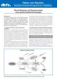
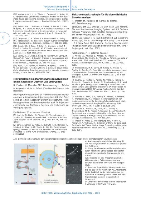
Abb. 1: Prinzip und Anwendung der elektronenspektroskopischen Abbildung (ESI) in der biomedizinischen Strukturanalyse.<br />
A: Strahlengang im analytischen Mikroskop für<br />
das Abbildungsverfahren mit inelastisch gestreuten<br />
Elektronen.<br />
B: Prinzip der elementarspezifischen Information<br />
durch inelastische Streuprozesse, bei welchen<br />
elementcharakteristische Energieverluste entstehen.<br />
C,D: Beispiele für eine Phosphor-spezifische<br />
Abbildung durch Elektronenspektroskopie<br />
simultan absorbierten TYMV und TMV Viruspartikeln.<br />
C: Typisches P-spezifisches Absorptionskantenbild<br />
bei 150 eV, der maximalen P-L 2,3 Absorptionskante.<br />
Zusätzlich zu der Information über die<br />
spezifische P-Verteilung enthält dieses Bild auch<br />
Signalbeiträge aus dem „unspezifischen“<br />
Untergrund.<br />
D: Differentiell errechnetes Bild der spezifischen<br />
P-Verteilung in TYMV und TMV Viren.